Medical Ultrasound Imaging
Sunday, 12 May 2024
Info Sheets Out- side     | 'Vascular Ultrasound' p3 Searchterm 'Vascular Ultrasound' found in 29 articles 3 terms [ • ] - 10 definitions [• ] - 16 booleans [• ]Result Pages : • Philips lacked in the field of ultrasound till 1998. By buying ATL (Bothell, Washington) in this year Philips establishing itself as an important player in ultrasound. In 2001 Philips also acquired Agilent (formerly Hewlett-Packard; Andover, Massachusetts), a market leader in the cardiology and vascular ultrasound systems (HP2000 to HP5500, now Sonos 2000 to Sonos 5500). Philips Medical System is the diagnostics business of Royal Philips Electronics of the Netherlands, one of the world's biggest electronics companies and Europe's largest. Philips is quoted on the NYSE (symbol: PHG), London, Frankfurt, Amsterdam and other stock exchanges. On October 19, 2001, Philips Medical Systems completed a 3-year acquisition strategy through its purchase of Marconi Medical Systems. Marconi Medical Systems offered leading multislice CT, MRI, and Nuclear Gamma Camera systems to medical institutions around the world. As well as new 3.0T developments, Philips is also in collaboration with researchers at the University of Nottingham, with the intention of developing an ultrahigh field strength clinical 7.0T whole body MR system. Ultrasound Systems:
•
•
•
•
•
(TEE) Transesophageal echocardiography provides a superior view of cardiac anatomy compared with transthoracic echocardiography. TEE is performed by the introduction of a probe attached to a fiberoptic endoscope into the esophagus. Caused by the position close to the heart e.g., clot finding and the view of the mitral valve are improved. Indications:
•
aortic atherosclerotic disease;
•
aortic dissection;
•
artificial mitral valves;
•
clots inside the left atrium;
•
cardiac infections;
•
masses or clots in the heart.
The piezoelectric crystal creating the acoustic power is mounted on the gastroscope that must be swallowed by the patient. This endoscopic transducer is miniaturized to approximately the size of a fingernail. Usually the probe is in place for an average of 15 minutes, to numb the surface a topical anesthetic is sprayed into the throat, in addition a conscious sedation is recommended. See also Myocardial Contrast Echocardiography, Stress Echocardiogram, M-Mode Echocardiography, Contrast Enhanced Ultrasound and Vascular Ultrasound Contrast Agents. • 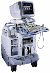 From GE Healthcare.;
'Vivid 7 Dimension, a premier cardiovascular ultrasound system from GE Healthcare, expands on the strength of a powerful imaging platform to offer new, innovative technology of dimensional proportions.'
Device Information and Specification
CONFIGURATION
Multi-frequency, linear, convex, phased, sector
B-mode, C-mode, M-mode (and 2-D), triplex mode, harmonic imaging, color flow mapping, 3D ultrasound display, power Doppler imaging (PDI), color Doppler, pulsed wave Doppler, continuous wave Doppler, tissue velocity imaging (TVI), tissue type imaging (TTI), strain rate imaging (SRI), tissue synchronization imaging (TSI)
IMAGING OPTIONS
CINE review with 5 speed types, bi- andtri-plane imaging with e.g. stress echo and tissue synchronization imaging
STORAGE, CONNECTIVITY, OS
Patient and image archive, HDD, MOD, DVD, USB flash card, DICOM 3.0 Windows-based
DATA PROCESSING
Digital beamformer with 1024 system processing channel technology
H*W*D m (inch.)
1.58 * 0.64 * 0.89 (62 * 25 * 35)
WEIGHT
191 kg (420 lbs.)
POWER CONSUMPTION
less than 2 KVA
• 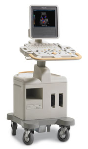 From Philips Medical Systems;
Introduced in June 2005, 'one of the less expensive and more dedicated' ultrasound systems.
Device Information and Specification
CONFIGURATION
LCD monitor
Broadband, convex, linear,
digital beamformer and focal tuning IMAGING OPTIONS
OPTIONAL PACKAGE
DICOM, etc.
STORAGE, CONNECTIVITY, OS
HDD, CD, USB, optionalMOD and DICOM 3.0
DATA PROCESSING
256-digitally processed channels
H*W*D inch.
58 * 20 * 32
WEIGHT
135 lbs.
• 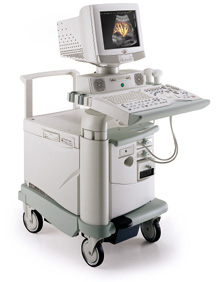 From Biosound Esaote, Inc.;
'Technos is an all-digital ultrasound system from Biosound Esaote, offering uncompromising diagnostic quality for Abdomen, Obstetric, Gynecology, Vascular, Breast, Small Parts and Urology applications. Experience the technological advantages of Technos, ... .' Specifications for this system will be available soon. Result Pages : | Share This Page Look Ups |
Medical-Ultrasound-Imaging.com
former US-TIP.com
Member of SoftWays' Medical Imaging Group - MR-TIP • Radiology TIP • Medical-Ultrasound-Imaging
Copyright © 2008 - 2024 SoftWays. All rights reserved.
Terms of Use | Privacy Policy | Advertise With Us
former US-TIP.com
Member of SoftWays' Medical Imaging Group - MR-TIP • Radiology TIP • Medical-Ultrasound-Imaging
Copyright © 2008 - 2024 SoftWays. All rights reserved.
Terms of Use | Privacy Policy | Advertise With Us
[last update: 2023-11-06 01:42:00]