Medical Ultrasound Imaging
Monday, 20 May 2024
Info Sheets Out- side     | 'Window' p2 Searchterm 'Window' found in 26 articles 6 terms [ • ] - 20 definitions [• ] Result Pages : •
The transforaminal or sub-occipital acoustic window is found in the space between the atlas and the base of the skull (through the foramen magnum
insonated from the top of the neck below the occiput). Practiced in the prone position (or sitting). This acoustic window allows the insonation of the vertebral arteries, basilar artery and some of the other branches of the posterior circulation (e.g. posterior inferior cerebellar artery). See also Transcranial Doppler. •
(TCCS) Transcranial color coded sonography is a combination of B-mode and pulsed wave Doppler. TCCS is used to study morphological and functional assessment of the circle of Willis, intracranial hemodynamics caused by extracranial artery stenosis, collateral flow and the vascular supply of intracranial lesion. Color imaging of the intracranial vessels allows placing the spectral Doppler volume correctly. This modality has encouraged the widespread use. Contrast enhanced TCCS analysis of cerebral arteriovenous transit time (cTT) is used as a measure of cerebral microcirculation. The windows that are used for transcranial Doppler examinations include regions where the skull bones are relatively thin or where naturally occurring gaps allow proper penetration of the sound beam. See also A-Mode, Cranial Bone Thermal Index, Transcranial Doppler and Transcranial Window. •
(TCD) Transcranial color Doppler sonography allows to evaluate the presence and flow direction of vessels as well as their relationships to surrounding structures. A disadvantage of cerebrovascular ultrasonography is the attenuation of the ultrasound signal by the skull. The loss of power through the skull is considerable, the signal to noise ratio is poor and so contrast enhanced Doppler imaging is advantageous. The use of ultrasound contrast agents provides a diagnostic window of sufficient duration and imaging quality to improve an evaluation of the cerebral vessels. Contrast TCD also results in visualization of small arteries and veins and greater length of these vessels. See also A-Mode, Cranial Bone Thermal Index, Transcranial Color Coded Sonography and Transcranial Window. Further Reading: Basics:
News & More:
•
Cerebrovascular ultrasonography is the best screening tool for the detection of carotid artery stenosis. Transcranial sonography is used in the evaluation of patients with suspected cerebrovascular disease, but a common problem is the attenuation of the ultrasound signal by the skull. Contrast enhanced ultrasound play a particularly important role in the visualization of the intracranial vessels, and thus improves the accuracy of transcranial Doppler and increases the potential of this technique. The use of microbubbles is helpful for classification of stenosis and for plaque evaluation in patients with poor initial examination. Ultrasound contrast agents avoid misdiagnosing a subtotal stenosis, which is a very important clinical issue. See also Adventitia, Intima, Periorbital Doppler, and Acoustic Window. • 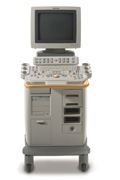 From Philips Medical Systems;
'Clinicians are demanding smaller, higher performing systems specifically designed to meet their clinical and operational challenges. The new Philips HD11 system provides an uncompromising platform, plus advanced options in a highly mobile and easy-to-use system.'
Device Information and Specification
APPLICATIONS
Abdominal, cardiac (also for adults with TEE), musculoskeletal (also pediatric), OB/GYN, prostate, smallparts, transcranial, vascular
CONFIGURATION
17' high resolution non-interlaced flat CRT, 4 active probe ports
B-mode, M-mode, coded harmonic imaging, color flow mode (CFM), power Doppler imaging (PDI), color Doppler, pulsed wave Doppler, tissue harmonic imaging
IMAGING OPTIONS
CrossXBeam spatial compounding, coded ultrasound acquisition),speckle reduction imaging (SRI), TruScan technology store raw data, CINE review with 4 speed types
OPTIONAL PACKAGE
Transesophageal scanning, stress echo, tissue velocity imaging (TVI), tissue velocity Doppler (TVD), contrast harmonic imaging
STORAGE, CONNECTIVITY, OS
Patient and image archive, HDD, DICOM 3.0, CD/DVD, MOD, Windows-based
DATA PROCESSING
Digital beamformer with 1024 system processing channel technology
H*W*D m (inch.)
1.62 * 0.61 * 0.99 (64 * 24 * 39)
WEIGHT
246 kg (498 lbs.)
POWER CONSUMPTION
less than 1.5 KVA
Result Pages : | Share This Page Look Ups |
Medical-Ultrasound-Imaging.com
former US-TIP.com
Member of SoftWays' Medical Imaging Group - MR-TIP • Radiology TIP • Medical-Ultrasound-Imaging
Copyright © 2008 - 2024 SoftWays. All rights reserved.
Terms of Use | Privacy Policy | Advertise With Us
former US-TIP.com
Member of SoftWays' Medical Imaging Group - MR-TIP • Radiology TIP • Medical-Ultrasound-Imaging
Copyright © 2008 - 2024 SoftWays. All rights reserved.
Terms of Use | Privacy Policy | Advertise With Us
[last update: 2023-11-06 01:42:00]