Medical Ultrasound Imaging
Wednesday, 8 May 2024
Info Sheets Out- side     | '3D Ultrasound' p4 Searchterm '3D Ultrasound' found in 23 articles 1 term [ • ] - 18 definitions [• ] - 4 booleans [• ]Result Pages : •
Unlike regular sound, ultrasound can be directed into a single direction. The echoes received by a stationary probe will result in a single dimensional signal showing peaks for every major material change. To generate a 2D picture, the probe is swiveled, either mechanically or through a phased array of ultrasound transducers. The data is analyzed by computer and used to construct the image. In a similar way, 3D pictures can be generated by computer using a specialized probe. In this way, a photo of an unborn baby may be made. Some ultrasonography machines can produce color pictures, of sorts. Doppler ultrasonography is color coded onto a gray scale picture. From the amount of energy in each echo, the difference in acoustic impedance can be calculated and a color is then assigned accordingly. See also Densitometry and 3D Ultrasound. •
Ultrasound technology with its advancements is vital for delivering high-quality patient care. Innovations including high-frequency ultrasound, 3D//4D imaging, contrast enhanced ultrasound, elastography, and point-of-care ultrasound, have expanded the capabilities of ultrasound imaging and improved diagnostic accuracy. B-Mode imaging, also known as brightness mode, is the fundamental technique in ultrasound imaging. It produces two-dimensional images based on the echoes received from tissues and organs. Understanding the principles of B-Mode imaging, such as gain adjustment, depth control, and image optimization, is crucial for obtaining diagnostically valuable images. M-Mode imaging, on the other hand, allows for the visualization of motion over time, enabling assessment of cardiac structures and function, as well as fetal heart rate. High-frequency ultrasound refers to the use of ultrasound waves with frequencies greater than 10 MHz. This technology enables improved resolution, allowing for detailed imaging of superficial structures like skin, tendons, and small organs. High-frequency ultrasound has found applications in dermatology, ophthalmology, and musculoskeletal imaging. Traditional 2D ultrasound has been augmented by the advent of 3D ultrasound technology. By acquiring multiple 2D images from different angles, this technique construct a volumetric representation of the imaged area. The addition of 4D ultrasound in real-time motion adds further value by capturing dynamic processes. Doppler imaging employs the Doppler effect to evaluate blood flow within vessels and assess hemodynamics. Color Doppler assigns color to different blood flow velocities, providing a visual representation of blood flow direction and speed. Spectral Doppler displays blood flow velocities as a waveform, allowing for detailed analysis of flow patterns, resistance, and stenosis. Contrast enhanced ultrasound employs microbubble contrast agents to enhance the visualization of blood flow and tissue perfusion. By injecting these agents intravenously, sonographers can differentiate between vascular structures and lesions. Elastography is a technique that measures tissue elasticity or stiffness. It assists in differentiating between normal and abnormal tissues, aiding in the diagnosis of various conditions such as liver fibrosis, breast lesions, and thyroid nodules. Fusion imaging combines ultrasound with other imaging modalities, such as computed tomography (CT), magnetic resonance imaging (MRI), or positron emission tomography (PET). By overlaying or merging ultrasound images with those obtained from other modalities, the user can precisely locate and characterize abnormalities, guide interventions, and improve diagnostic accuracy. Fusion imaging has proven particularly useful in areas such as interventional radiology, oncology, and urology. See also Equipment Preparation, Environmental Protection, Handheld Ultrasound, Portable Ultrasound and Ultrasound Accessories and Supplies. •
Conventional, CT and MR imaging technologies are limited in their availability, to depict soft tissue, or to show dynamic activity, like cardiac muscle contractility and blood flow. Easy applicability, real-time sonography and biopsy facilitation are important advantages in veterinarian medicine. Veterinary ultrasound has a very high sensitivity to show the composition of soft tissues, but the low specificity is a disadvantage. High ultrasound system performance includes Doppler techniques, contrast enhanced ultrasound, 3D ultrasound, and tissue harmonic imaging to improve resolution. Technical and physical requirements of veterinary ultrasound are the same as in human ultrasonography. The higher the sound frequency, the better the possible resolution, but the poorer the tissue penetration. Image quality is depended of the ultrasound equipment. For example, a 10 MHz transducer is excellent for imaging of superficial structures; a 3.5 or 5.0 megahertz transducer allows sufficient penetration to see inner structures like the liver or the heart. In addition, the preparation and performing of the examination is similar to that of humans. The sound beam penetrates soft tissue and fat well, but gas and bone impede the ultrasonic power. Fluid filled organs like the bladder are often used as an acoustic window, and an ultrasound gel is used to conduct the sound beam. • 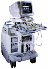 From GE Healthcare.;
'Vivid 7 Dimension, a premier cardiovascular ultrasound system from GE Healthcare, expands on the strength of a powerful imaging platform to offer new, innovative technology of dimensional proportions.'
Device Information and Specification
CONFIGURATION
Multi-frequency, linear, convex, phased, sector
B-mode, C-mode, M-mode (and 2-D), triplex mode, harmonic imaging, color flow mapping, 3D ultrasound display, power Doppler imaging (PDI), color Doppler, pulsed wave Doppler, continuous wave Doppler, tissue velocity imaging (TVI), tissue type imaging (TTI), strain rate imaging (SRI), tissue synchronization imaging (TSI)
IMAGING OPTIONS
CINE review with 5 speed types, bi- andtri-plane imaging with e.g. stress echo and tissue synchronization imaging
STORAGE, CONNECTIVITY, OS
Patient and image archive, HDD, MOD, DVD, USB flash card, DICOM 3.0 Windows-based
DATA PROCESSING
Digital beamformer with 1024 system processing channel technology
H*W*D m (inch.)
1.58 * 0.64 * 0.89 (62 * 25 * 35)
WEIGHT
191 kg (420 lbs.)
POWER CONSUMPTION
less than 2 KVA
• 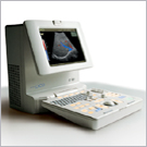 From Medison Co.,Ltd.;
'Full-featured Digital Imaging On Your Desktop The SONOACE PICO is a full-featured digital color ultrasound system with virtually all the imaging capabilities you would expect from a cart-based system with the added benefit of being fully portable. Built on a reliable Linux PC platform, the SONOACE PICO boasts all- digital beamforming and signal processing for best-in-class image resolution enhanced by a long list of advanced diagnostic tools such as harmonic imaging, color and power Doppler, and freehand 3D imaging.' Result Pages : | Share This Page Look Ups |
Medical-Ultrasound-Imaging.com
former US-TIP.com
Member of SoftWays' Medical Imaging Group - MR-TIP • Radiology TIP • Medical-Ultrasound-Imaging
Copyright © 2008 - 2024 SoftWays. All rights reserved.
Terms of Use | Privacy Policy | Advertise With Us
former US-TIP.com
Member of SoftWays' Medical Imaging Group - MR-TIP • Radiology TIP • Medical-Ultrasound-Imaging
Copyright © 2008 - 2024 SoftWays. All rights reserved.
Terms of Use | Privacy Policy | Advertise With Us
[last update: 2023-11-06 01:42:00]