Medical Ultrasound Imaging
Wednesday, 8 May 2024
Info Sheets Out- side     | 'Abdominal Ultrasound' p2 Searchterm 'Abdominal Ultrasound' found in 24 articles 1 term [ • ] - 6 definitions [• ] - 17 booleans [• ]Result Pages : •
As far as ultrasound is concerned, 4D ultrasound (also referred to as live 3D ultrasound or 4B-mode) is the latest ultrasound technology - the fourth dimension means length, width, and depth over time. 4D Ultrasound takes 3D ultrasound images and adds the element of time to the progress so that a moving three-dimensional image is seen on the monitor. A 4D scan takes the same amounts of time as a 2D or 3D scan; the difference is the ultrasound equipment being used. One advantage of a 4D fetal ultrasound to a 2D-mode is that parents can see how their baby will generally look like. However, there are different opinions over the medical advantages. To scan a 3D ultrasound image, the probe is swept over the maternal abdomen. A computer takes multiple images and renders the 3D picture. With 4D imaging, the computer takes the images as multiple pictures while the probe is hold still and a 3D image is simultaneously rendered in real time on a monitor. In most cases, the standard 2D ultrasound is taken, and then the 3D/4D scan capability is added if an abnormality is detected or suspected. The 3D/4D sonogram is then focused on a specific area, to provide the details needed to assess and diagnose a suspected problem. A quick 4D scan of the face of the fetus may be performed at the end of a routine exam, providing the parents with a photo. See also Obstetric and Gynecologic Ultrasound, Pregnancy Ultrasound, Fetal Ultrasound and Abdominal Ultrasound. •
Diagnostic ultrasound imaging has no known risks or long-term side effects. Discomfort to the patient is very rare if the sonogram is accurately performed by using appropriate frequencies and intensity ranges. However, the application of the ALARA principle is always recommended.
There are reports of low birth weight of babies after applying more than the recommended ultrasound examinations during pregnancy. Women who think they might be pregnant should raise this issue with the doctor before undergoing an abdominal ultrasound, to avoid any harm to the fetus in the early stages of development. Since ultrasound is energy, sensitive tissues like the reproductive organs could possibly sustain damage if vibrated to a high degree by too intense ultrasound waves. In diagnostic ultrasonic procedures, such damage would only result from improper use of the equipment. Possible ultrasound bioeffects:
•
•
Due to increasing of temperature, dissolved gases from microbubbles come out of the contrast solution.
The thermal effect is controlled by the displayed thermal index and the mechanical index indicates the risk of cavitation. An ultrasound gel is applied to obtain better contact between the transducer and the skin. This has the consistency of thick mineral oil and is not associated with skin irritation or allergy. Specific conditions for which ultrasound may be selected as a treatment may be attached with higher risks. See also Ultrasound Imaging Procedures, Fetal Ultrasound and Obstetric and Gynecologic Ultrasound. • 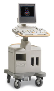 From Philips Medical Systems;
Introduced in June 2005, 'one of the less expensive and more dedicated' ultrasound systems.
Device Information and Specification
CONFIGURATION
LCD monitor
Broadband, convex, linear,
digital beamformer and focal tuning IMAGING OPTIONS
OPTIONAL PACKAGE
DICOM, etc.
STORAGE, CONNECTIVITY, OS
HDD, CD, USB, optionalMOD and DICOM 3.0
DATA PROCESSING
256-digitally processed channels
H*W*D inch.
58 * 20 * 32
WEIGHT
135 lbs.
•  From GE Healthcare.;
'GE is defining a new age of ultrasound. We call it Volume Ultrasound. GE's Voluson 730 Expert is a powerful system that enables real-time techniques for acquiring, navigating and analyzing volumetric images so that you can make clinical decisions with unprecedented confidence.'
Device Information and Specification
APPLICATIONS
Abdominal, breast, cardiac, musculoskeletal, neonatal, OB/GYN, pediatric, small parts, transcranial, urological, vascular
CONFIGURATION
15' high resolution non-interlaced flat CRT, 4 active probe ports
B-mode, M-mode, coded harmonic imaging (2-D), color flow mode (CFM), power Doppler imaging (PDI), color Doppler, pulsed wave Doppler, high pulse repetition frequency (HPRF) Doppler, tissue harmonic imaging, 3-D power Doppler
IMAGING OPTIONS
CrossXBeam spatial compounding, coded excitation , spatio-temporal image correlation (STIC), B-Flow (simultaneous imaging of tissue and blood flow), strain rate imaging (SRI)
OPTIONAL PACKAGE
STORAGE, CONNECTIVITY, OS
SonoView archiving and data management, network, HDD, DICOM 3.0, CD/DVD, MOD, USB, Windows-based
DATA PROCESSING
Digital beamformer with 512 system processing channel technology
H*W*D m (inch.)
1.43 * 0.69 * 1.02 (56 * 27 * 40)
WEIGHT
136 kg (300 lbs.)
•  From GE Healthcare.;
'Vivid 7 Dimension, a premier cardiovascular ultrasound system from GE Healthcare, expands on the strength of a powerful imaging platform to offer new, innovative technology of dimensional proportions.'
Device Information and Specification
CONFIGURATION
Multi-frequency, linear, convex, phased, sector
B-mode, C-mode, M-mode (and 2-D), triplex mode, harmonic imaging, color flow mapping, 3D ultrasound display, power Doppler imaging (PDI), color Doppler, pulsed wave Doppler, continuous wave Doppler, tissue velocity imaging (TVI), tissue type imaging (TTI), strain rate imaging (SRI), tissue synchronization imaging (TSI)
IMAGING OPTIONS
CINE review with 5 speed types, bi- andtri-plane imaging with e.g. stress echo and tissue synchronization imaging
STORAGE, CONNECTIVITY, OS
Patient and image archive, HDD, MOD, DVD, USB flash card, DICOM 3.0 Windows-based
DATA PROCESSING
Digital beamformer with 1024 system processing channel technology
H*W*D m (inch.)
1.58 * 0.64 * 0.89 (62 * 25 * 35)
WEIGHT
191 kg (420 lbs.)
POWER CONSUMPTION
less than 2 KVA
Result Pages : | Share This Page Look Ups |
Medical-Ultrasound-Imaging.com
former US-TIP.com
Member of SoftWays' Medical Imaging Group - MR-TIP • Radiology TIP • Medical-Ultrasound-Imaging
Copyright © 2008 - 2024 SoftWays. All rights reserved.
Terms of Use | Privacy Policy | Advertise With Us
former US-TIP.com
Member of SoftWays' Medical Imaging Group - MR-TIP • Radiology TIP • Medical-Ultrasound-Imaging
Copyright © 2008 - 2024 SoftWays. All rights reserved.
Terms of Use | Privacy Policy | Advertise With Us
[last update: 2023-11-06 01:42:00]