Medical Ultrasound Imaging
Sunday, 20 September 2026

 Info Sheets  Out- side     | 'Mode' p14 Searchterm 'Mode' found in 134 articles 14 terms [ • ] - 120 definitions [• ] Result Pages : • 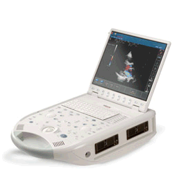 From ESAOTE S.p.A.;
'The MyLab™30CV ultrasound system is an evolutionary step in ultrasound technology. Weighing less than 20 pounds, it is the first compact ultrasound system to deliver premium console performance. And with mobile, portable or stationary configurations, MyLab30CV can adapt to any clinical environment.'
Device Information and Specification
APPLICATIONS
Abdominal, breast, cardiac, OB/GYN, pediatric, pediatric cardiology, small parts, transcranial, vascular
CONFIGURATION
Portable
Linear: 4-10 MHz, convex: 2-5 MHz, phased: 1.6-10 MHz, micro convex: 5-7.5 MHz, endocavity: 5-7.5 MHz, pencil: 2 + 5 MHz
2-D, M-mode, duplex, triplex, color Doppler, pulsed wave Doppler, tissue velocity mapping (TVM), tissue enhancement imaging (TEI™), contrast harmonic imaging, stress echo, tissue velocity mapping for LV motion analysis (TVM), contrast tuned imaging for contrast media procedures (CnTI™), Qontrast™ for myocardium parameters quantification
STORAGE, CONNECTIVITY, OS
Digital patient archive/management, integrated CD/RW, RJ 45 and USB ports, Windows
H*W*D m (inch.)
0.16 * 0.36 * 0.50 (6.2 x 14 x 19.3)
WEIGHT
Less than 11 kg (20 lbs.)
•
As far as ultrasound is concerned, 4D ultrasound (also referred to as live 3D ultrasound or 4B-mode) is the latest ultrasound technology - the fourth dimension means length, width, and depth over time. 4D Ultrasound takes 3D ultrasound images and adds the element of time to the progress so that a moving three-dimensional image is seen on the monitor. A 4D scan takes the same amounts of time as a 2D or 3D scan; the difference is the ultrasound equipment being used. One advantage of a 4D fetal ultrasound to a 2D-mode is that parents can see how their baby will generally look like. However, there are different opinions over the medical advantages. To scan a 3D ultrasound image, the probe is swept over the maternal abdomen. A computer takes multiple images and renders the 3D picture. With 4D imaging, the computer takes the images as multiple pictures while the probe is hold still and a 3D image is simultaneously rendered in real time on a monitor. In most cases, the standard 2D ultrasound is taken, and then the 3D/4D scan capability is added if an abnormality is detected or suspected. The 3D/4D sonogram is then focused on a specific area, to provide the details needed to assess and diagnose a suspected problem. A quick 4D scan of the face of the fetus may be performed at the end of a routine exam, providing the parents with a photo. See also Obstetric and Gynecologic Ultrasound, Pregnancy Ultrasound, Fetal Ultrasound and Abdominal Ultrasound. •
Ultrasound technology has evolved significantly, providing sonographers with a wide range of ultrasound machines. As technology has advanced, portable ultrasound equipment, including handheld ultrasound systems, have emerged in the field of medical imaging. However, these devices may have limited imaging capabilities and reduced image quality compared to larger systems. Types of ultrasound systems compiled according to their portability: •
Handheld Ultrasound Devices: Handheld ultrasound devices are compact, lightweight, and easily maneuverable. They offer convenience and point-of-care imaging capabilities, making them ideal for emergency medicine, primary care, and remote settings. Pros include portability, rapid assessments, and ease of use. However, these devices may have limited imaging capabilities and reduced image quality compared to larger systems. •
Laptop-Based Ultrasound Scanner: Laptop-based ultrasound machines combine portability with a larger display and enhanced imaging capabilities. They are versatile and suitable for various applications, including primary care, obstetrics, and musculoskeletal imaging. These machines provide good image quality, a user-friendly interface, and improved storage capacity. However, they may still be bulkier and less portable than handheld devices. •
Console-Based Ultrasound Systems:
Console-based ultrasound machines are larger, stationary systems commonly found in hospitals and specialized imaging centers. They offer comprehensive imaging capabilities, advanced features, and excellent image quality. These machines are suitable for a wide range of specialties and can perform complex examinations. Pros include high image resolution, advanced imaging modes, and comprehensive data management. However, they lack the portability and immediate accessibility of handheld or laptop-based devices.
•
Cart-Based Ultrasound Machines: Cart-based ultrasound machines strike a balance between portability and advanced imaging capabilities. They consist of a console unit mounted on a mobile cart. These machines are commonly used in hospitals, clinics, and larger healthcare facilities. They provide excellent image quality, a wide range of imaging options, and ergonomic considerations. While less portable than handheld or laptop-based devices, cart-based machines offer enhanced functionality and versatility. In summary, pros and cons of portable ultrasound machines: •
Pros: Compact portable ultrasound machines eliminate transfers and reduce wait times, improving patient comfort. Scans at the bedside minimize discomfort and anxiety while optimizing workflow efficiency. Sonographers can quickly assess patients and detect abnormalities. Real-time examinations provide immediate visualization, procedural guidance, and support for critical decision-making especially in emergency, critical care, and resource-limited settings. •
Cons: Portable ultrasound machines may have restricted features and imaging modes compared to larger systems, potentially affecting diagnostic quality and detail. The compact size can lead to compromises in image resolution and overall quality due to factors like lower power output and smaller transducers.The small displays and simplified controls of portable systems may pose challenges for sonographers, potentially impacting workflow efficiency and user fatigue. Portable ultrasound machines often come with a higher price tag, requiring careful evaluation of cost versus expected benefits and specific practice needs. See also Ultrasound Accessories and Supplies, Environmental Protection, Sonographer, Ultrasound Technology and Equipment Preparation.  Further Reading: News & More:
•
The thermal effect of ultrasound is caused by absorption of the ultrasound beam energy. As the ultrasound waves are absorbed, their energy is converted into heat. The higher the frequency, the greater the absorbed dose, converted to heat according the equation: f = 1/T where T is the period as in simple harmonic motion. Ultrasound is a mechanical energy in which a pressure wave travels through tissue. Heat is produced at the transducer surface and also tissue in the depth can be heated as ultrasound is absorbed. The thermal effect is highest in tissue with a high absorption coefficient, particularly in bone, and is low where there is little absorption. The temperature rise is also dependent on the thermal characteristics of the tissue (conduction of heat and perfusion), the ultrasound intensity and the length of examination time. The intensity is also dependent on the power output and the position of the tissue in the beam profile. The intensity at a particular point can be changed by many of the operator controls, for example power output, mode (B-mode, color flow, spectral Doppler), scan depth, focus, zoom and area of color flow imaging. The transducer face and tissue in contact with the transducer can be heated. See also Thermal Units Per Hour and Ultrasound Radiation Force. •
(TCCS) Transcranial color coded sonography is a combination of B-mode and pulsed wave Doppler. TCCS is used to study morphological and functional assessment of the circle of Willis, intracranial hemodynamics caused by extracranial artery stenosis, collateral flow and the vascular supply of intracranial lesion. Color imaging of the intracranial vessels allows placing the spectral Doppler volume correctly. This modality has encouraged the widespread use. Contrast enhanced TCCS analysis of cerebral arteriovenous transit time (cTT) is used as a measure of cerebral microcirculation. The windows that are used for transcranial Doppler examinations include regions where the skull bones are relatively thin or where naturally occurring gaps allow proper penetration of the sound beam. See also A-Mode, Cranial Bone Thermal Index, Transcranial Doppler and Transcranial Window. Result Pages : | Share This Page     Look Ups |
 (.pdf)
Medical-Ultrasound-Imaging.com
former US-TIP.com
Member of SoftWays' Medical Imaging Group - MR-TIP • Radiology TIP • Medical-Ultrasound-Imaging
Copyright © 2008 - 2025 SoftWays. All rights reserved.
Terms of Use | Privacy Policy | Advertise With Us
former US-TIP.com
Member of SoftWays' Medical Imaging Group - MR-TIP • Radiology TIP • Medical-Ultrasound-Imaging
Copyright © 2008 - 2025 SoftWays. All rights reserved.
Terms of Use | Privacy Policy | Advertise With Us
[last update: 2025-04-05 02:38:00]