Medical Ultrasound Imaging
Thursday, 31 October 2024
'Power Map' Searchterm 'Power Map' found in 8 articles 1 term [ • ] - 2 definitions [• ] - 5 booleans [• ]Result Pages : • •
Color saturation is a characteristic used for example to describe the vividness of a color, or the gradation of hue from unsaturated (white) to fully saturated (100% of the given color). See also Directional Indicators, and Power Map. •
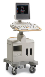
In power mode the amplitude (power) of color Doppler signals is displayed, regardless of the velocity. Power does not have negative values and is independent of sampling frequency. An aliasing artifact does not occur in power mode images. Caused by plotting the quantity enhanced by echo contrast agents in a power map, power mode is often used in contrast Doppler ultrasound examinations. Also known as energy mode. •  From Philips Medical Systems;
Introduced in June 2005, 'one of the less expensive and more dedicated' ultrasound systems.
Device Information and Specification
CONFIGURATION
LCD monitor
Broadband, convex, linear,
digital beamformer and focal tuning IMAGING OPTIONS
OPTIONAL PACKAGE
DICOM, etc.
STORAGE, CONNECTIVITY, OS
HDD, CD, USB, optionalMOD and DICOM 3.0
DATA PROCESSING
256-digitally processed channels
H*W*D inch.
58 * 20 * 32
WEIGHT
135 lbs.
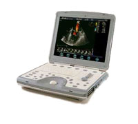
•  From GE Healthcare.;
'The incredible Vivid i system establishes a completely new level of cardiovascular performance that gives clinicians the freedom to get diagnostic results outside of the echo lab.'
Device Information and Specification
APPLICATIONS
CONFIGURATION
Notebook
M-mode (and 2-D), triplex mode, harmonic imaging, color flow mapping, pulsed wave Doppler, continuous wave Doppler, power Doppler, color Doppler, tissue harmonic imaging, color flow mapping
IMAGING OPTIONS
STORAGE, CONNECTIVITY, OS
Patient and image archive, HDD, DICOM, CD/DVD, MOD, USB flash, PCMCIA, eVue for remote monitoring, MPEGvue foruniversal record sharing
H*W*D cm (inch.)
7 * 36 * 32 (2.6 x 14.1 x 12.3)
WEIGHT
5 kg (11 lbs.)
POWER CONSUMPTION
Rechargeable battery provides up to 1.0 hour of full scan operation
Result Pages : |
Medical-Ultrasound-Imaging.com
former US-TIP.com
Member of SoftWays' Medical Imaging Group - MR-TIP • Radiology TIP • Medical-Ultrasound-Imaging
Copyright © 2008 - 2024 SoftWays. All rights reserved.
Terms of Use | Privacy Policy | Advertise With Us
former US-TIP.com
Member of SoftWays' Medical Imaging Group - MR-TIP • Radiology TIP • Medical-Ultrasound-Imaging
Copyright © 2008 - 2024 SoftWays. All rights reserved.
Terms of Use | Privacy Policy | Advertise With Us
[last update: 2023-11-06 01:42:00]


