Medical Ultrasound Imaging
Wednesday, 8 May 2024
Info Sheets Out- side     | '4D Ultrasound' p3 Searchterm '4D Ultrasound' found in 15 articles 1 term [ • ] - 10 definitions [• ] - 4 booleans [• ]Result Pages : •
Ultrasound technology with its advancements is vital for delivering high-quality patient care. Innovations including high-frequency ultrasound, 3D//4D imaging, contrast enhanced ultrasound, elastography, and point-of-care ultrasound, have expanded the capabilities of ultrasound imaging and improved diagnostic accuracy. B-Mode imaging, also known as brightness mode, is the fundamental technique in ultrasound imaging. It produces two-dimensional images based on the echoes received from tissues and organs. Understanding the principles of B-Mode imaging, such as gain adjustment, depth control, and image optimization, is crucial for obtaining diagnostically valuable images. M-Mode imaging, on the other hand, allows for the visualization of motion over time, enabling assessment of cardiac structures and function, as well as fetal heart rate. High-frequency ultrasound refers to the use of ultrasound waves with frequencies greater than 10 MHz. This technology enables improved resolution, allowing for detailed imaging of superficial structures like skin, tendons, and small organs. High-frequency ultrasound has found applications in dermatology, ophthalmology, and musculoskeletal imaging. Traditional 2D ultrasound has been augmented by the advent of 3D ultrasound technology. By acquiring multiple 2D images from different angles, this technique construct a volumetric representation of the imaged area. The addition of 4D ultrasound in real-time motion adds further value by capturing dynamic processes. Doppler imaging employs the Doppler effect to evaluate blood flow within vessels and assess hemodynamics. Color Doppler assigns color to different blood flow velocities, providing a visual representation of blood flow direction and speed. Spectral Doppler displays blood flow velocities as a waveform, allowing for detailed analysis of flow patterns, resistance, and stenosis. Contrast enhanced ultrasound employs microbubble contrast agents to enhance the visualization of blood flow and tissue perfusion. By injecting these agents intravenously, sonographers can differentiate between vascular structures and lesions. Elastography is a technique that measures tissue elasticity or stiffness. It assists in differentiating between normal and abnormal tissues, aiding in the diagnosis of various conditions such as liver fibrosis, breast lesions, and thyroid nodules. Fusion imaging combines ultrasound with other imaging modalities, such as computed tomography (CT), magnetic resonance imaging (MRI), or positron emission tomography (PET). By overlaying or merging ultrasound images with those obtained from other modalities, the user can precisely locate and characterize abnormalities, guide interventions, and improve diagnostic accuracy. Fusion imaging has proven particularly useful in areas such as interventional radiology, oncology, and urology. See also Equipment Preparation, Environmental Protection, Handheld Ultrasound, Portable Ultrasound and Ultrasound Accessories and Supplies. •  From GE Healthcare.;
'GE is defining a new age of ultrasound. We call it Volume Ultrasound. GE's Voluson 730 Expert is a powerful system that enables real-time techniques for acquiring, navigating and analyzing volumetric images so that you can make clinical decisions with unprecedented confidence.'
Device Information and Specification
APPLICATIONS
Abdominal, breast, cardiac, musculoskeletal, neonatal, OB/GYN, pediatric, small parts, transcranial, urological, vascular
CONFIGURATION
15' high resolution non-interlaced flat CRT, 4 active probe ports
B-mode, M-mode, coded harmonic imaging (2-D), color flow mode (CFM), power Doppler imaging (PDI), color Doppler, pulsed wave Doppler, high pulse repetition frequency (HPRF) Doppler, tissue harmonic imaging, 3-D power Doppler
IMAGING OPTIONS
CrossXBeam spatial compounding, coded excitation , spatio-temporal image correlation (STIC), B-Flow (simultaneous imaging of tissue and blood flow), strain rate imaging (SRI)
OPTIONAL PACKAGE
STORAGE, CONNECTIVITY, OS
SonoView archiving and data management, network, HDD, DICOM 3.0, CD/DVD, MOD, USB, Windows-based
DATA PROCESSING
Digital beamformer with 512 system processing channel technology
H*W*D m (inch.)
1.43 * 0.69 * 1.02 (56 * 27 * 40)
WEIGHT
136 kg (300 lbs.)
• 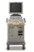 From Philips Medical Systems;
'Clinicians are demanding smaller, higher performing systems specifically designed to meet their clinical and operational challenges. The new Philips HD11 system provides an uncompromising platform, plus advanced options in a highly mobile and easy-to-use system.'
Device Information and Specification
APPLICATIONS
Abdominal, cardiac (also for adults with TEE), musculoskeletal (also pediatric), OB/GYN, prostate, smallparts, transcranial, vascular
CONFIGURATION
17' high resolution non-interlaced flat CRT, 4 active probe ports
B-mode, M-mode, coded harmonic imaging, color flow mode (CFM), power Doppler imaging (PDI), color Doppler, pulsed wave Doppler, tissue harmonic imaging
IMAGING OPTIONS
CrossXBeam spatial compounding, coded ultrasound acquisition),speckle reduction imaging (SRI), TruScan technology store raw data, CINE review with 4 speed types
OPTIONAL PACKAGE
Transesophageal scanning, stress echo, tissue velocity imaging (TVI), tissue velocity Doppler (TVD), contrast harmonic imaging
STORAGE, CONNECTIVITY, OS
Patient and image archive, HDD, DICOM 3.0, CD/DVD, MOD, Windows-based
DATA PROCESSING
Digital beamformer with 1024 system processing channel technology
H*W*D m (inch.)
1.62 * 0.61 * 0.99 (64 * 24 * 39)
WEIGHT
246 kg (498 lbs.)
POWER CONSUMPTION
less than 1.5 KVA
•  From Philips Medical Systems;
'The Philips iU22 system combines Intelligent Design, including breakthroughs in ergonomics, with Intelligent Control, providing new levels of automation, to give you revolutionary performance and workflow.'
Device Information and Specification
APPLICATIONS
Abdominal, cardiac (also for adults with TEE), musculoskeletal (also pediatric), OB/GYN, prostate, smallparts, transcranial, vascular
CONFIGURATION
17' high resolution non-interlaced flat CRT, 4 active probe ports
B-mode, M-mode, coded harmonic imaging, color flow mode (CFM), power Doppler imaging (PDI), color Doppler, pulsed wave Doppler, tissue harmonic imaging
IMAGING OPTIONS
CrossXBeam spatial compounding, coded ultrasound acquisition),speckle reduction imaging (SRI), TruScan technology store raw data, CINE review with 4 speed types
OPTIONAL PACKAGE
Transesophageal scanning, stress echo, tissue velocity imaging (TVI), tissue velocity Doppler (TVD), contrast harmonic imaging
STORAGE, CONNECTIVITY, OS
Patient and image archive, HDD, DICOM 3.0, CD/DVD, MOD, Windows-based
DATA PROCESSING
Digital beamformer with 1024 system processing channel technology
H*W*D m (inch.)
1.62 * 0.61 * 0.99 (64 * 22 * 43)
WEIGHT
kg (345 lbs.)
POWER CONSUMPTION
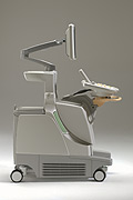
•  From Siemens Medical Systems;
'This high-performance, multi-specialty system supports and improves your daily ultrasound routine. Embedded DICOM creates the integrated foundation for a complete connectivity solution while MultiHertz™ multiple frequency imaging and Tissue Harmonic Imaging (THI) expands the clinical versatility of the system. Advanced applications such as stress echo, SieScape™ panoramic imaging, and transesophageal imaging can be seamlessly integrated.'
Device Information and Specification
CLINICAL APPLICATION
Widest range of applications
CONFIGURATION
Compact, mobile system
Wideband MultiHertz™ multiple frequency
IMAGING OPTIONS
OPTIONAL PACKAGE
Software upgradeability to advanced clinical application
IMAGING ENHANCEMENTS
Precision MotionCapture, Synthetic aperture technology
STORAGE
Patient and image database management system
DATA PROCESSING
Parallel and quad signal processing
WEIGHT
Lightweight
Result Pages : | Share This Page Look Ups |
Medical-Ultrasound-Imaging.com
former US-TIP.com
Member of SoftWays' Medical Imaging Group - MR-TIP • Radiology TIP • Medical-Ultrasound-Imaging
Copyright © 2008 - 2024 SoftWays. All rights reserved.
Terms of Use | Privacy Policy | Advertise With Us
former US-TIP.com
Member of SoftWays' Medical Imaging Group - MR-TIP • Radiology TIP • Medical-Ultrasound-Imaging
Copyright © 2008 - 2024 SoftWays. All rights reserved.
Terms of Use | Privacy Policy | Advertise With Us
[last update: 2023-11-06 01:42:00]