Medical Ultrasound Imaging
Wednesday, 15 May 2024
Info Sheets Out- side     | 'Bit' p3 Searchterm 'Bit' found in 23 articles 3 terms [ • ] - 20 definitions [• ] Result Pages : •
(CWD) Continuous wave (CW) Doppler is an ultrasound imaging mode, which records blood flow velocities along the length of the beam. Continuous wave Doppler uses different crystals to send and receive the signal. The transducer operating in continuous wave mode utilizes one half of the elements and is continuously sending sound waves of a single frequency while the other half is continuously receiving the reflected signals. The advantages of a continuous wave transducer are a high sensitivity and no Nyquist limit. CW Doppler does not alias but has no depth precision and large gate. The beat frequency is the Doppler shift. CW Doppler echocardiography employs this technique to record the flow of blood through the cardiovascular system. See also Cross Talk, Periorbital Doppler, and Mirror Artifact. Further Reading: News & More:
•  From Lantheus Medical Imaging.
Activated DEFINITY® Injectable Suspension is indicated for use in patients with suboptimal echocardiograms to opacify the left ventricular chamber and to improve the delineation of the left ventricular endocardial border. The perflutren lipid microspheres exhibit lower acoustic impedance than blood and enhance the intrinsic backscatter of blood. Echocardiography with Definity produced more detailed images of the heart in difficult-to-image patients versus echocardiography alone, and images obtained with the contrast agent provided a more accurate assessment of segmental wall motion compared with unenhanced images. See also Coherent Contrast Imaging. In August 2001 DuPont Pharmaceuticals Company received FDA approval.
Drug Information and Specification
RESEARCH NAME
DMP 115, MRX 115
INDICATION -
DEVELOPMENT STAGE APPLICATION
Intravenous
TYPE
Lipids:DPPA, DPPC,MPEG5000 DPPE
CHARGE
Negative
Octafluoropropane
MICROBUBBLE SIZE
98% < 10μm
PRESENTATION
Package contains four 2mL clear glass single-use vials.
STORAGE
Refrigerate 2−8 °C
PREPARATION
Activate through Vialmix agitation
DO NOT RELY ON THE INFORMATION PROVIDED HERE, THEY ARE
NOT A SUBSTITUTE FOR THE ACCOMPANYING PACKAGE INSERT!
Distribution Information
TERRITORY
DISTRIBUTOR
North America, Australia, South Asia, Middle East
Further Reading: Basics:
News & More:
•
Environmental protection in ultrasound imaging involves adopting practices and technologies that minimize the environmental impact associated with the use of ultrasound equipment and disposables. Here are some key considerations: •
Energy Efficiency: Opt for energy-efficient ultrasound machines and equipment that are designed to minimize energy consumption. This helps reduce the overall environmental impact associated with power usage. •
Digitalization and Paper Reduction: Embrace digital imaging and archiving systems to reduce reliance on paper. Storing images and reports electronically minimizes paper consumption, printing supplies, and physical storage space. •
Waste Management: Implement proper waste management practices for ultrasound-related disposables, such as ultrasound gel bottles, probe covers, and cleaning materials. Follow local regulations for the disposal of medical waste and prioritize recycling and responsible disposal methods. •
Equipment Lifespan and Disposal: Choose ultrasound equipment known for its durability and longevity. Maximizing the lifespan of equipment reduces the frequency of replacements, minimizing electronic waste generation. When disposing of old equipment, ensure proper recycling and disposal in accordance with local regulations. •
Education and Awareness: Promote education and awareness among ultrasound professionals about environmentally conscious practices. Encourage staff to adopt energy-saving habits, such as turning off equipment when not in use, and emphasize the importance of responsible waste management. Develop standardized and optimized examination protocols to minimize the duration and number of ultrasound scans required per patient. This helps reduce the energy consumption associated with prolonged imaging sessions and decreases the overall environmental impact. By focusing on energy efficiency, digitalization, waste management, equipment lifespan, and education, healthcare facilities can make significant strides towards reducing their carbon footprint and the environmental impact of ultrasound imaging practices. See also Ultrasound System Performance, Equipment Preparation, Ultrasound Accessories and Supplies and Sonographer. •
Equipment Preparation is an essential step in ensuring optimal ultrasound imaging quality and maintaining a safe and hygienic scanning environment. The following considerations should be taken into account:
•
Ultrasound Machine Warm-Up: The ultrasound scanner should be turned on and allowed to warm up for at least 5 minutes before initiating the examination. This allows the system to stabilize and ensures consistent performance. •
Transducer Selection: The appropriate pobe should be selected based on the type of examination required, as well as the patient's body size, weight, and habitus. Different transducer offer varying frequencies, field of view, and imaging capabilities, allowing for tailored imaging based on the specific clinical needs. •
Power Settings and Techniques: Prior to beginning the examination, it is crucial to verify and adjust the power settings and imaging techniques according to the examination protocol. This ensures that the ultrasound machine is optimized for the specific diagnostic requirements •
Acoustic Couplant Application: An adequate amount of acoustic couplant, such as warmed ultrasound gel, should be applied to the patient's skin or the transducer surface. This gel serves as a medium that promotes maximum transmission of the sound beam by eliminating air interfaces, leading to improved image quality. •
Transducer Cleaning and Probe Covers: All transducers should be cleaned and readily available for use with each patient. While endocavitary ultrasound probes are often protected by single-use disposable probe covers, it is important to maintain proper hygiene by performing a high-level disinfection of the probe between each use. Additionally, using a probe cover as an additional measure can help keep the probe clean and minimize the risk of cross-contamination. By following these equipment preparation guidelines, healthcare professionals can ensure accurate and safe ultrasound examinations while promoting infection control measures and maintaining a hygienic environment for both patients and staff. See also Environmental Protection, Portable Ultrasound Machine, Ultrasound Accessories and Supplies, and Ultrasound System Performance. • 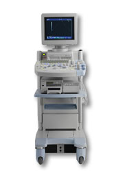 From Hitachi Medical Corporation (HMC), sales, marketing and service in the US by Hitachi Medical Systems America Inc.
The HI VISION™ 5500 - EUB-5500 fully digital ultrasound system delivers the latest generation of signal processing technology, sophisticated transducer design, and a host of features and options for advanced imaging capabilities across a wide range of clinical situations. This system is compatible with all Pentax ultrasound endoscopes.
Device Information and Specification
APPLICATIONS
Abdominal, brachytherapy/cryotherapy, breast, cardiac, dedicated biopsy, endoscopic, intraoperative, laparoscopic, musculoskeletal, OB/GYN, pediatric, small parts, urologic, vascular
CONFIGURATION
Compact system
Five frequency (except mini-probes)
Linear, convex, radial, miniradial/miniprobe, biplane, phased array, echoendoscope longitudinal, echoendoscope radial
3 modes of dynamic tissue harmonic imaging (dTHI), pulsed wave Doppler, continuous wave Doppler, color flow imaging, power Doppler, directional power Doppler, color flow angiography, real-time Doppler measurements
IMAGING OPTIONS
3RD generation color artifact suppression
OPTIONAL PACKAGE
STORAGE, CONNECTIVITY, OS
Patient and image database management system, HDD, FDD, MOD, CD-ROM, Network, DICOM 3.0, Windows XP
DATA PROCESSING
H*W*D m (inch.)
1.40 x 0.51 x 0.79 (55 x 20 x 31)
WEIGHT
130 kg (286 lbs.)
POWER CONSUMPTION
1.2kVA
ENVIRONMENTAL IMPACT
4096 btu/hr heat output
Result Pages : | Share This Page Look Ups |
Medical-Ultrasound-Imaging.com
former US-TIP.com
Member of SoftWays' Medical Imaging Group - MR-TIP • Radiology TIP • Medical-Ultrasound-Imaging
Copyright © 2008 - 2024 SoftWays. All rights reserved.
Terms of Use | Privacy Policy | Advertise With Us
former US-TIP.com
Member of SoftWays' Medical Imaging Group - MR-TIP • Radiology TIP • Medical-Ultrasound-Imaging
Copyright © 2008 - 2024 SoftWays. All rights reserved.
Terms of Use | Privacy Policy | Advertise With Us
[last update: 2023-11-06 01:42:00]