Medical Ultrasound Imaging
Friday, 10 May 2024
Info Sheets Out- side     | 'Contrast Resolution' p2 Searchterm 'Contrast Resolution' found in 23 articles 7 definitions [ • ] - 16 booleans [• ]Result Pages : •
Tissue-specific ultrasound contrast agents improve the image contrast resolution through differential uptake. The concentration of microbubble contrast agents within the vasculature, reticulo-endothelial, or lymphatic systems produces an effective passive targeting of these areas. Other contrast media concepts include targeted drug delivery via contrast microbubbles. Tissue-specific ultrasound contrast agents are injected intravenously and taken up by specific tissues or they adhere to specific targets such as venous thrombosis. These effects may require minutes to several hours to reach maximum effectiveness. By enhancing the acoustic differences between normal and diseased tissues, these tissue-specific agents improve the detectability of abnormalities. Some microbubbles accumulate in normal hepatic tissue; some are phagocytosed by Kupffer cells in the reticuloendothelial system and others may stay in the sinusoids. Liver tumors without normal Kupffer cells can be identified by the lack of the typical mosaic color pattern of the induced acoustic emission. The hepatic parenchymal phase, which may last from less than an hour to several days, depending on the specific contrast medium used, may be imaged by bubble-specific modes such as stimulated acoustic emission (color Doppler using high MI) or pulse inversion imaging. Further Reading: News & More:
•
Ultrasound machines, with their various components and types, have revolutionized the field of medical imaging. These devices enable healthcare professionals to visualize internal structures, assess conditions, and guide interventions with real-time imaging capabilities.
Today, medical ultrasound systems are complex signal processing machines. Assessing the performance of an ultrasound system requires understanding the relationships between the characteristics of the system, such as the point spread function, temporal resolution, and the quality of images. Image quality aspects include the detail resolution, contrast resolution and penetration. Systems with microbubble scanner modification are particularly suitable for contrast enhanced ultrasound.
•
Low-performance systems constitute approximately 20% of the world ultrasound market. These ultrasound machines are characterized by basic black and white imaging and are primarily used for basic OB/GYN applications and fetal development monitoring. They are often purchased by private office practitioners and small hospitals, with a unit cost below $50,000. These scanners commonly come equipped with a transvaginal probe.
•
Mid-performance sonography systems also hold around 20% market share. These machines are basic gray scale imaging, color and spectral Doppler and are used for routine examinations and reporting. They typically utilize a minimum number of scanheads and find applications in radiology, cardiology, and OB/GYN. The cost of these systems ranges between $50,000 and $100,000. Refurbished advanced and high-performance ultrasound machines with fewer optional features can also be found in this price range.
•
High-performance ultrasound systems generally provide high-resolution gray scale imaging, advanced color power and spectral Doppler capabilities. They usually include advanced measurement and analysis software, image review capabilities, and a variety of probes. These high-performance sonography devices have a market share of approximately 40% and cost between $100,000 and $150,000.
•
The remaining 20% of the market consists of premium or advanced performance ultrasound systems, typically sold for over $150,000. Premium performance systems offer high-resolution gray scale imaging, advanced color flow, power Doppler, and spectral Doppler, as well as features like tissue harmonic imaging, image acquisition storage, display and review capabilities, advanced automation, and more. Premium systems are equipped with a wide assortment of transducer scanheads.
In summary, ultrasound machines have diverse performance levels and corresponding price ranges, catering to various medical imaging needs. From low-performance systems with basic imaging capabilities to high-performance and premium systems with advanced features, ultrasound technology continues to advance healthcare imaging capabilities. See also Ultrasound Physics, Handheld Ultrasound, Environmental Protection, Equipment Preparation. Further Reading: Basics:
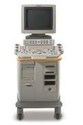
•  From GE Healthcare.;
'GE is defining a new age of ultrasound. We call it Volume Ultrasound. GE's Voluson 730 Expert is a powerful system that enables real-time techniques for acquiring, navigating and analyzing volumetric images so that you can make clinical decisions with unprecedented confidence.'
Device Information and Specification
APPLICATIONS
Abdominal, breast, cardiac, musculoskeletal, neonatal, OB/GYN, pediatric, small parts, transcranial, urological, vascular
CONFIGURATION
15' high resolution non-interlaced flat CRT, 4 active probe ports
B-mode, M-mode, coded harmonic imaging (2-D), color flow mode (CFM), power Doppler imaging (PDI), color Doppler, pulsed wave Doppler, high pulse repetition frequency (HPRF) Doppler, tissue harmonic imaging, 3-D power Doppler
IMAGING OPTIONS
CrossXBeam spatial compounding, coded excitation , spatio-temporal image correlation (STIC), B-Flow (simultaneous imaging of tissue and blood flow), strain rate imaging (SRI)
OPTIONAL PACKAGE
STORAGE, CONNECTIVITY, OS
SonoView archiving and data management, network, HDD, DICOM 3.0, CD/DVD, MOD, USB, Windows-based
DATA PROCESSING
Digital beamformer with 512 system processing channel technology
H*W*D m (inch.)
1.43 * 0.69 * 1.02 (56 * 27 * 40)
WEIGHT
136 kg (300 lbs.)
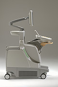
•  From Philips Medical Systems;
'Clinicians are demanding smaller, higher performing systems specifically designed to meet their clinical and operational challenges. The new Philips HD11 system provides an uncompromising platform, plus advanced options in a highly mobile and easy-to-use system.'
Device Information and Specification
APPLICATIONS
Abdominal, cardiac (also for adults with TEE), musculoskeletal (also pediatric), OB/GYN, prostate, smallparts, transcranial, vascular
CONFIGURATION
17' high resolution non-interlaced flat CRT, 4 active probe ports
B-mode, M-mode, coded harmonic imaging, color flow mode (CFM), power Doppler imaging (PDI), color Doppler, pulsed wave Doppler, tissue harmonic imaging
IMAGING OPTIONS
CrossXBeam spatial compounding, coded ultrasound acquisition),speckle reduction imaging (SRI), TruScan technology store raw data, CINE review with 4 speed types
OPTIONAL PACKAGE
Transesophageal scanning, stress echo, tissue velocity imaging (TVI), tissue velocity Doppler (TVD), contrast harmonic imaging
STORAGE, CONNECTIVITY, OS
Patient and image archive, HDD, DICOM 3.0, CD/DVD, MOD, Windows-based
DATA PROCESSING
Digital beamformer with 1024 system processing channel technology
H*W*D m (inch.)
1.62 * 0.61 * 0.99 (64 * 24 * 39)
WEIGHT
246 kg (498 lbs.)
POWER CONSUMPTION
less than 1.5 KVA
•  From Philips Medical Systems;
'The Philips iU22 system combines Intelligent Design, including breakthroughs in ergonomics, with Intelligent Control, providing new levels of automation, to give you revolutionary performance and workflow.'
Device Information and Specification
APPLICATIONS
Abdominal, cardiac (also for adults with TEE), musculoskeletal (also pediatric), OB/GYN, prostate, smallparts, transcranial, vascular
CONFIGURATION
17' high resolution non-interlaced flat CRT, 4 active probe ports
B-mode, M-mode, coded harmonic imaging, color flow mode (CFM), power Doppler imaging (PDI), color Doppler, pulsed wave Doppler, tissue harmonic imaging
IMAGING OPTIONS
CrossXBeam spatial compounding, coded ultrasound acquisition),speckle reduction imaging (SRI), TruScan technology store raw data, CINE review with 4 speed types
OPTIONAL PACKAGE
Transesophageal scanning, stress echo, tissue velocity imaging (TVI), tissue velocity Doppler (TVD), contrast harmonic imaging
STORAGE, CONNECTIVITY, OS
Patient and image archive, HDD, DICOM 3.0, CD/DVD, MOD, Windows-based
DATA PROCESSING
Digital beamformer with 1024 system processing channel technology
H*W*D m (inch.)
1.62 * 0.61 * 0.99 (64 * 22 * 43)
WEIGHT
kg (345 lbs.)
POWER CONSUMPTION
Result Pages : | Share This Page Look Ups |
Medical-Ultrasound-Imaging.com
former US-TIP.com
Member of SoftWays' Medical Imaging Group - MR-TIP • Radiology TIP • Medical-Ultrasound-Imaging
Copyright © 2008 - 2024 SoftWays. All rights reserved.
Terms of Use | Privacy Policy | Advertise With Us
former US-TIP.com
Member of SoftWays' Medical Imaging Group - MR-TIP • Radiology TIP • Medical-Ultrasound-Imaging
Copyright © 2008 - 2024 SoftWays. All rights reserved.
Terms of Use | Privacy Policy | Advertise With Us
[last update: 2023-11-06 01:42:00]