Medical Ultrasound Imaging
Thursday, 9 May 2024
Info Sheets Out- side     | 'Dynamic Range' p2 Searchterm 'Dynamic Range' found in 15 articles 1 term [ • ] - 4 definitions [• ] - 10 booleans [• ]Result Pages : •
Linear array transducer elements are rectangular and arranged in a line. Linear array probes are described by the radius of width in mm. A linear array transducer can have up to 512 elements spaced over 75-120 mm. The beam produced by such a narrow element will diverge rapidly after the wave travels only a few millimeters. The smaller the face of the transducer, the more divergent is the beam. This would result in poor lateral resolution due to beam divergence and low sensitivity due to the small element size. In order to overcome this, adjacent elements are pulsed simultaneously (typically 8 to 16; or more in wide-aperture designs). In a subgroup of x elements, the inner elements pulse delayed with respect to the outer elements. The interference of the x small divergent wavelets produces a focused beam. The delay time determines the depth of focus for the transmitted beam and can be changed during scanning. Linear arrays are usually cheaper than sector scanners but have greater skin contact and therefore make it difficult to reach organs between ribs such as the heart. One-dimensional linear array transducers may have dynamic, electronic focusing providing a narrow ultrasound beam in the image plane. In the z-plane (elevation plane - perpendicular to the image plane) focusing may be provided by an acoustic lens with a fixed focal zone. Rectangular or matrix transducers with unequal rows of transducer elements are two-dimensional (2D), but they are termed 1.5D, because the number of rows is much less than the number of columns. These transducers provide dynamic, electronic focusing even in the z-plane. See also Rectangular Array Transducer. •
Transducers used for the real-time mode are different than for the A-mode, B-, or M-modes. A linear array transducer with multiple piezoelectric crystal elements that are different arranged and fired, transmits the needed larger sound beam. A subgroup of x adjacent elements (8-16; or more in wide-aperture designs) is pulsed simultaneously; the inner elements pulse delayed with respect to the outer elements. The interference of the x small divergent wavelets generates a focused beam. The delay time determining the focus depth of a real-time transducer can be changed during imaging. Similar delay factors applied during the receiving phase, result in a dynamic focusing effect on the return. This forms a single scan line in the real-time image. To produce the following scan line, another group of x elements is selected by shifting one element position along the transducer array from the previous group. This pattern is then repeated for the groups along the array, in a sequential and repetitive way. Further Reading: Basics: • 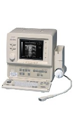 From ALOKA Co., Ltd.;
'A High-Quality, Portable Veterinary Ultrasound System. The SSD-500V is a rugged, portable convex sector/linear scanner that offers versatility, superb image quality and reliable performance in a compact design. This 22 lb system has Electronic dynamic focus and 256 shades of gray that produces high-quality images. The SSD-500V includes comprehensive measurement and analysis packages. Measurements include distance/depth, area, circumference, velocity, heart rate, ratio and angle. The system also included obstetrical analysis functions that can calculate estimated gestational age and expected date of confinement. In addition, customized obstetrical tables can be easily created. The SSD-500V software package also includes left ventricular function software for M-mode Cardiac evaluation. The 500V provides excellent clinical versatility − for use in both small and large animal applications − using a wide range of optional convex and linear array transducers.' • 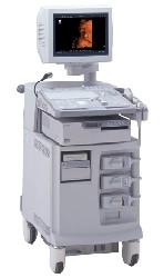 From ALOKA Co., Ltd.;
'The ProSound SSD-4000 utilizes the most advanced acoustic technologies available today, and its multidisciplinary technology architecture enables it to offer great versatility and flexibility over a wide range of clinical applications. With its new-generation, front-end technology including a 12-bit A/D converter, the ProSound SSD-4000 offers superior contrast resolution−especially when compared to 10-bit systems.'
Device Information and Specification
APPLICATIONS
CONFIGURATION
Compact, portable, dual dynamic display
Wide-band super high-density (W-SHD) transducers
OPTIONAL PACKAGE
Volume Mode
STORAGE, CONNECTIVITY, OS
Data Management Subsystem (iDMS), DICOM-Worklist
DATA PROCESSING
•
From Bayer Schering Pharma AG: Echovist-200® was an effectively one-pass-only contrast medium for contrast sonography and Doppler-echocardiographic examinations for the detection, exclusion or follow-up of pathological states leading to hemodynamic changes. Because of the short intravascular life of the microparticles and microbubbles, transit through the pulmonary circulation is unusual. In cardiac evaluations Echovist-200® has been replaced by newer ultrasound contrast agents (USCA), therefore the manufacturing was discontinued. Another range of echo contrast application is the female genital tract, in particular for the demonstration or exclusion of acquired or congenital changes of the uterine cavity and for the visualization of the Fallopian tubes and investigation of their patency. 1 g Echovist-200 granules contain 1 g D-galactose microparticles. 1 ml aqueous solution for production of the suspension contains 200 mg D-galactose. Brand names in other countries: Ecovist.
Drug Information and Specification
RESEARCH NAME
-
DEVELOPER
INDICATION
Hysterosalpingo-contrast sonography (HyCoSy), echocardiographic use in neonates and children
APPLICATION
Intravenous injection
TYPE
Microbubble
D-GALACTOSE®
Air
MICROBUBBLE SIZE
99 % < 12 μm, 95 % < 8 μm
STORAGE
Store below 30 °C
PRESENTATION
Vials of 20 ml with 3.0 g granulate incl. one vial of 15 ml containing 13.5 ml D-galactose solution, one mini-spike
PREPARATION
Reconstitute with water
DO NOT RELY ON THE INFORMATION PROVIDED HERE, THEY ARE
NOT A SUBSTITUTE FOR THE ACCOMPANYING PACKAGE INSERT! Further Reading: News & More:
Result Pages : | Share This Page Look Ups |
Medical-Ultrasound-Imaging.com
former US-TIP.com
Member of SoftWays' Medical Imaging Group - MR-TIP • Radiology TIP • Medical-Ultrasound-Imaging
Copyright © 2008 - 2024 SoftWays. All rights reserved.
Terms of Use | Privacy Policy | Advertise With Us
former US-TIP.com
Member of SoftWays' Medical Imaging Group - MR-TIP • Radiology TIP • Medical-Ultrasound-Imaging
Copyright © 2008 - 2024 SoftWays. All rights reserved.
Terms of Use | Privacy Policy | Advertise With Us
[last update: 2023-11-06 01:42:00]