Medical Ultrasound Imaging
Sunday, 19 May 2024
Info Sheets Out- side     | 'Harmonic Imaging' p4 Searchterm 'Harmonic Imaging' found in 44 articles 6 terms [ • ] - 38 definitions [• ] Result Pages : • 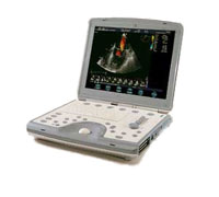 From GE Healthcare.;
'The incredible Vivid i system establishes a completely new level of cardiovascular performance that gives clinicians the freedom to get diagnostic results outside of the echo lab.'
Device Information and Specification
APPLICATIONS
CONFIGURATION
Notebook
M-mode (and 2-D), triplex mode, harmonic imaging, color flow mapping, pulsed wave Doppler, continuous wave Doppler, power Doppler, color Doppler, tissue harmonic imaging, color flow mapping
IMAGING OPTIONS
STORAGE, CONNECTIVITY, OS
Patient and image archive, HDD, DICOM, CD/DVD, MOD, USB flash, PCMCIA, eVue for remote monitoring, MPEGvue foruniversal record sharing
H*W*D cm (inch.)
7 * 36 * 32 (2.6 x 14.1 x 12.3)
WEIGHT
5 kg (11 lbs.)
POWER CONSUMPTION
Rechargeable battery provides up to 1.0 hour of full scan operation
• View NEWS results for 'Vivid i' (1). •  From GE Healthcare.;
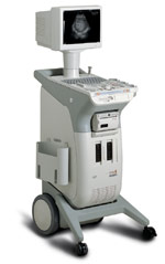
'GE is defining a new age of ultrasound. We call it Volume Ultrasound. GE's Voluson 730 Expert is a powerful system that enables real-time techniques for acquiring, navigating and analyzing volumetric images so that you can make clinical decisions with unprecedented confidence.'
Device Information and Specification
APPLICATIONS
Abdominal, breast, cardiac, musculoskeletal, neonatal, OB/GYN, pediatric, small parts, transcranial, urological, vascular
CONFIGURATION
15' high resolution non-interlaced flat CRT, 4 active probe ports
B-mode, M-mode, coded harmonic imaging (2-D), color flow mode (CFM), power Doppler imaging (PDI), color Doppler, pulsed wave Doppler, high pulse repetition frequency (HPRF) Doppler, tissue harmonic imaging, 3-D power Doppler
IMAGING OPTIONS
CrossXBeam spatial compounding, coded excitation , spatio-temporal image correlation (STIC), B-Flow (simultaneous imaging of tissue and blood flow), strain rate imaging (SRI)
OPTIONAL PACKAGE
STORAGE, CONNECTIVITY, OS
SonoView archiving and data management, network, HDD, DICOM 3.0, CD/DVD, MOD, USB, Windows-based
DATA PROCESSING
Digital beamformer with 512 system processing channel technology
H*W*D m (inch.)
1.43 * 0.69 * 1.02 (56 * 27 * 40)
WEIGHT
136 kg (300 lbs.)
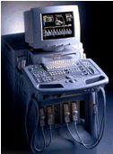
•  From Medison Co.,Ltd.;
'The Ultimate Black & White Ultrasound System. Redefining the standard of excellence in digital B/W ultrasound. Medison, the innovator in medical science, proudly introduces the ultimate evolution in black-and-white ultrasound system - the 128 BW. With its revolutionary responsive imaging technology, powered by Medison's harmonic imaging and digital 128 channel beamforming, 128 BW produces crystal-clear images in fraction of time. Designed and committed for excellent performance, 128 BW guarantees its scientific breakthrough results.' •
In 3D ultrasound (US) several 2D images are acquired by moving the probe across the body surface or rotating inserted probes. 3D-mode uses the same basic concept of a 2D ultrasound but rather than take the image from a single angle, the sonographer takes a volume image. The volume image that is displayed on the screen is a software rendering of all of the detected soft-tissue combined by specialized computer software to form three-dimensional images. The 3D volume rendering technique (VR) does not rely on segmentation (segmentation techniques are difficult to apply to ultrasound pictures) and makes it possible to obtain clear 3D ultrasound images for clinical diagnosis. A 3D ultrasound produces a still image. Diagnostic US systems with 3D display functions and linear array probes are mainly used for obstetric and abdominal applications. The combination of contrast agents, harmonic imaging and power Doppler greatly improves 3D US reconstructions. 3D imaging shows a better look at the organ being examined and is used for:
•
Detection of abnormal fetus development, e.g. of the face and limbs.
•
Visualization of e.g. the colon and rectum.
•
Pictures of blood flow in various organs or a fetus.
Fusion 3D imaging methods for generating compound images from two sets of ultrasound images (B-mode and Doppler images) enable the observation of the structural relationships between lesions and their associated blood vessels in three dimensions (maximum intensity projection). Further Reading: News & More:
•  From Siemens Medical Systems;
The new gold standard in echocardiography is defined by Native Tissue Harmonic Imaging (NTHI) and DELTA Differential Echo Amplification−two powerful technologies that were first introduced on Acuson's Sequoia™ echocardiography system. Now they have migrated to the Aspen™ Advanced system. Specifications for this system will be available soon. Result Pages : | Share This Page Look Ups |
Medical-Ultrasound-Imaging.com
former US-TIP.com
Member of SoftWays' Medical Imaging Group - MR-TIP • Radiology TIP • Medical-Ultrasound-Imaging
Copyright © 2008 - 2024 SoftWays. All rights reserved.
Terms of Use | Privacy Policy | Advertise With Us
former US-TIP.com
Member of SoftWays' Medical Imaging Group - MR-TIP • Radiology TIP • Medical-Ultrasound-Imaging
Copyright © 2008 - 2024 SoftWays. All rights reserved.
Terms of Use | Privacy Policy | Advertise With Us
[last update: 2023-11-06 01:42:00]