Medical Ultrasound Imaging
Thursday, 9 May 2024
Info Sheets Out- side     | 'Out of Phase' p3 Searchterm 'Out of Phase' found in 19 articles 1 definition [ • ] - 18 booleans [• ]Result Pages : •
Quadrature detection is used in Doppler ultrasound as well as in magnetic resonance imaging and is also called quadrature demodulation or phase quadrature technique. Quadrature detection is the acquisition of Mx and My simultaneously as a function of time by using two separate detector channels. This signal processing method is used for directional Doppler in which the signal reference frequency for the two channels has a phase shift of 1/4 period. The output Doppler signal phase for both channels also depends on the Doppler shift whether positive or negative. The fast Fourier transform analyzer performs spectral Doppler analysis in ultrasound machines and displays different quadrature Doppler frequencies, when a sample volume cursor is used along time. •
(UCA / USCA) Ultrasonography is the most commonly performed diagnostic imaging procedure. The introduction of sonographic contrast media into routine practice modifies the use of ultrasound in a variety of clinical applications. USCAs consist of microbubbles filled with air or gases and can be classified according to their pharmacokinetics. Among the blood pool agents, transpulmonary ultrasound contrast agents offer higher diagnostic potential compared to agents that cannot pass the pulmonary capillary bed after a peripheral intravenous injection. In addition to their vascular phase, some USCAs can exhibit a tissue- or organ-specific phase. The sonogram image quality is improved either by decreasing the reflectivity of the undesired interfaces or by increasing the backscattered echoes from the desired regions. Different types of ultrasound contrast agents: Ultrasound contrast agents act as echo-enhancers, because of the high different acoustic impedance at the interface between gas and blood. The enhanced echo intensity is proportional to the change in acoustical impedance as the sound beam crosses from the blood to the gas in the bubbles. The ideal qualities of an ultrasound contrast agent:
•
high echogenicity;
•
low attenuation;
•
low blood solubility;
•
low diffusivity;
•
ability to pass through the pulmonary capillary bed;
•
lack of biological effects with repeat doses.
A typical ultrasound contrast agent consists of a thin flexible or rigid shell composed of albumin, lipid, or polymer confining a gas such as nitrogen, or a perfluorocarbon. The choice of the microbubble shell and gas has an important influence on the properties of the agent. Current generations of microbubbles have a diameter from 1 μm to 5 μm. The success of these agents is mostly dependent on the small size and on the stability of their shell, which allows passage of the microbubbles through the pulmonary circulation. Microbubbles must be made smaller than the diameter of capillaries or they would embolize and be ineffective and perhaps even dangerous. The reflectivity of these microbubbles is proportional to the fourth power of a particle diameter but also directly proportional to the concentration of the contrast agent particles themselves. Ultrasound contrast agents produce unique acoustic signatures that allow to separate their signal from tissue echoes and to depict whether they are moving or stationary. This enables the detection of capillary flow and of targeted microbubbles that are retained in tissues such as normal liver. The new generation of contrast media is characterized by prolonged persistence in the vascular bed which provides consistent enhancement of the arterial Doppler signal. Contrast agents make it also possible to perform dynamic and perfusion studies. Targeted contrast imaging agents are for example taken up by the phagocytic cell systems and thus have liver/spleen specific effects. See also Ultrasound Contrast Agent Safety, Adverse Reaction, Tissue-Specific Ultrasound Contrast Agent, and Bubble Specific Imaging. Further Reading: Basics:
News & More:
•
A bolus is a rapid infusion of high dose contrast agent. Dynamic and accumulation phase imaging can be performed after bolus injection. Since the transit time of the bolus is only a short time, images with high frame rate show the wash in and wash out of the contrast material.
The injection rate and the total injected volume modifies the bolus peak profile. Substantial changes in the concentrations during signal acquisition induce artifacts. Furthermore, the hemodynamic parameters (cardiac output, blood pressure) influence the bolus profile. However, the characteristics of ultrasound contrast agents are favorable with a continuous perfusion. See also Negative Bolus. • 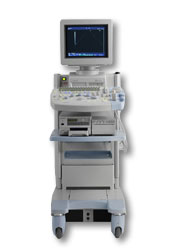 From Hitachi Medical Corporation (HMC), sales, marketing and service in the US by Hitachi Medical Systems America Inc.
The HI VISION™ 5500 - EUB-5500 fully digital ultrasound system delivers the latest generation of signal processing technology, sophisticated transducer design, and a host of features and options for advanced imaging capabilities across a wide range of clinical situations. This system is compatible with all Pentax ultrasound endoscopes.
Device Information and Specification
APPLICATIONS
Abdominal, brachytherapy/cryotherapy, breast, cardiac, dedicated biopsy, endoscopic, intraoperative, laparoscopic, musculoskeletal, OB/GYN, pediatric, small parts, urologic, vascular
CONFIGURATION
Compact system
Five frequency (except mini-probes)
Linear, convex, radial, miniradial/miniprobe, biplane, phased array, echoendoscope longitudinal, echoendoscope radial
3 modes of dynamic tissue harmonic imaging (dTHI), pulsed wave Doppler, continuous wave Doppler, color flow imaging, power Doppler, directional power Doppler, color flow angiography, real-time Doppler measurements
IMAGING OPTIONS
3RD generation color artifact suppression
OPTIONAL PACKAGE
STORAGE, CONNECTIVITY, OS
Patient and image database management system, HDD, FDD, MOD, CD-ROM, Network, DICOM 3.0, Windows XP
DATA PROCESSING
H*W*D m (inch.)
1.40 x 0.51 x 0.79 (55 x 20 x 31)
WEIGHT
130 kg (286 lbs.)
POWER CONSUMPTION
1.2kVA
ENVIRONMENTAL IMPACT
4096 btu/hr heat output
•  From Hitachi Medical Corporation (HMC);
The HI VISION™ 6500 - EUB-6500 high resolution digital ultrasound system offers advanced clinical imaging, enhanced operating efficiency, and remarkable clinical flexibility, all in robust and versatile configuration that simply represents a better clinical solution in a variety of real-world, real-work arenas.
Device Information and Specification
APPLICATIONS
Abdominal, brachytherapy/cryotherapy, breast, cardiac, dedicated biopsy, endoscopic, intraoperative, laparoscopic, musculoskeletal, OB/GYN, pediatric, small parts, urologic, vascular
CONFIGURATION
Compact system
Linear, convex, radial, miniradial/miniprobe, biplane, phased array, echoendoscope longitudinal, echoendoscope radial
Tissue Doppler imaging (TDI), pulsed wave Doppler, continuous wave Doppler, color flow imaging, power Doppler, directional power Doppler, color flow angiography, real-time Doppler measurements, 4 modes of dynamic tissue harmonic imaging (dTHI), wideband pulse inversion imaging (WPI)
IMAGING OPTIONS
3RD generation color artifact suppression
OPTIONAL PACKAGE
3D ultrasound, dual omni-directional M-mode display, steerable CW Doppler, dynamic contrast harmonics imaging, stress echo, Pentax EUS and Fujinon Mini-probe
STORAGE, CONNECTIVITY, OS
Patient and image database management system, HDD, FDD, MOD, CD-ROM, Network, DICOM 3.0, Windows XP
DATA PROCESSING
H*W*D m (inch.)
1.40 x 0.51 x 0.79 (55 x 20 x 31)
WEIGHT
130 kg (286 lbs.)
POWER CONSUMPTION
1.2kVA
ENVIRONMENTAL POLLUTION
4096 btu/hr heat output
Result Pages : | Share This Page Look Ups |
Medical-Ultrasound-Imaging.com
former US-TIP.com
Member of SoftWays' Medical Imaging Group - MR-TIP • Radiology TIP • Medical-Ultrasound-Imaging
Copyright © 2008 - 2024 SoftWays. All rights reserved.
Terms of Use | Privacy Policy | Advertise With Us
former US-TIP.com
Member of SoftWays' Medical Imaging Group - MR-TIP • Radiology TIP • Medical-Ultrasound-Imaging
Copyright © 2008 - 2024 SoftWays. All rights reserved.
Terms of Use | Privacy Policy | Advertise With Us
[last update: 2023-11-06 01:42:00]