Medical Ultrasound Imaging
Sunday, 19 May 2024
Info Sheets Out- side     | 'Pancreas Ultrasound' p2 Searchterm 'Pancreas Ultrasound' found in 11 articles 11 booleans [ • ]Result Pages : •
Sonography [aka: ultrasonography] is a term that encompasses the entire process of performing ultrasound examinations and interpreting the obtained images. Sonography involves the skilled application of ultrasound technology by trained professionals known as sonographers or ultrasound technologists. These specialists operate the ultrasound equipment, manipulate the transducer, and acquire the necessary pictures for diagnostic imaging purposes. Sonography requires in-depth knowledge of anatomy, physiology, and pathology to accurately interpret the ultrasound images and provide valuable information to the treating physician. Sonography uses equipment that generates high frequency sound waves to produce images from muscles, soft tissues, fluid collections, and vascular structures of the human body. Obstetric sonography is commonly used during pregnancy. Sonography visualizes anatomy, function, and pathology of for example gallbladder, kidneys, pancreas, spleen, liver, uterus, ovaries, urinary bladder, eye, thyroid, breast, aorta, veins and arteries in the extremities, carotid arteries in the neck, as well as the heart. A typical medical ultrasound machine, usually a real-time scanner, operates in the frequency range of 2 to 13 megahertz. See also Musculoskeletal and Joint Ultrasound, Pediatric Ultrasound, Cerebrovascular Ultrasonography and Contrast Enhanced Ultrasound. Further Reading: Basics:
News & More:
•
(AUS) Abdominal ultrasound, also known as abdominal sonography, is a medical imaging technique that focuses on the visualization and assessment of the abdominal organs. While 'abdominal ultrasound' is the commonly used term, there are alternative terms that can be used to refer to this imaging modality: (TAE) transabdominal echography, abdominal ultrasonography, sonogram, FAST (Focused Assessment with Sonography for Trauma). Abdominal ultrasound imaging is an invaluable clinical tool for identifying the underlying cause of abdominal pain. An abdominal ultrasound examination encompasses a comprehensive evaluation of the liver, gallbladder, biliary tree, pancreas, spleen, kidneys, and abdominal blood vessels. It is a cost-effective, safe, and non-invasive medical imaging modality that is typically utilized as the initial diagnostic investigation. Advanced ultrasound techniques, such as high-resolution ultrasound, endoscopic ultrasound, and contrast-enhanced Doppler, further enhance the detection of small lesions and provide detailed information for precise diagnosis. To prepare for an abdominal ultrasound, it is recommended to have nothing to eat or drink for at least 8 hours, starting from midnight the night before the examination. Indications:
•
Abdominal pain
•
Gallbladder or kidneys stones
•
Inflammation
•
Detection of cancer and metastasis
FAST (Focused Assessment with Sonography for Trauma) is a rapid diagnostic test used for trauma patients. It sequentially evaluates the presence of free fluid in the pericardium (hemopericardium) and in four specific views of the abdomen. These views include the right upper quadrant (RUQ), left upper quadrant (LUQ), subcostal, and suprapubic views. They aid in identifying hemoperitoneum in patients with potential truncal injuries. The space between the liver and the right kidney (RUQ), known as Morison's pouch, is a location where intraperitoneal fluid can accumulate. Emergency abdominal ultrasonography is indicated in cases of suspected aortic aneurysm, appendicitis, biliary and renal colic, as well as blunt or penetrating abdominal trauma. It plays a crucial role in the timely assessment and management of these conditions, providing critical information to guide appropriate treatment decisions. See also Handheld Ultrasound, Pelvic Ultrasound, Pregnancy Ultrasound, Prostate Ultrasound, Interventional Ultrasound and Pediatric Ultrasound. Further Reading: Basics: News & More:
• 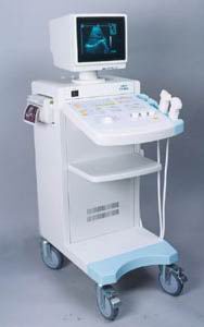 From SIUI Inc.;
'The CTS-310B is designed for the diagnosis of liver, gall, kidney, pancreas, thyroid, breast, uterus, bladder, ovary, etc. It is a versatile ultrasound scanner with both linear array scanning and convex scanning.' Features: 'Powerful image processing circuit & High quality image A wide range of probes for selection Cineloop Probe frequency conversation option Computer image communication Various measuring function Backlit keyboard'
Device Information and Specification
APPLICATIONS
See description above
CONFIGURATION
Normal system, 10' high resolution monitor, dual probe connector
Linear and convex
PROBES STANDARD
2.5MHz to 10.0MHz, linear and convex, broad band, trifrequency
IMAGING OPTIONS
Multi zoom rate and depth shift
OPTIONAL PACKAGE
POWER REQUIREMENT
AC 220V/110V, 50Hz/60Hz
POWER CONSUMPTION
0.1 KVA
• 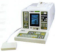 From SIUI Inc.;
'The SIUI CTS-200 is one of few 'linear only' units to be developed with a digital scan converter to process all incoming signals. With a digital processor, not only is the incoming signal processed faster but less noise is introduced into the signal path. The images are cleaner, crisper than you have experienced, even in machines at many times the price.' 'CTS-200 is suitable to a wide range of examination of liver, gallbladder, kidney, spleen, pancreas, thyroid gland, breast, uterus, urinary bladder, OB/GYN, etc. It is a portable ultrasound scanner of high performance.'
Device Information and Specification
APPLICATIONS
See description above
CONFIGURATION
Portable, gray scale(256)
Linear
PROBES STANDARD
1 * 3.5MHz Linear Probe EZU-PL21
PROBES FREQUENCY
3.5MHz, 7.5MHz
IMAGING OPTIONS
*1, *1.5, *2 as well as depth shift
OPTIONAL PACKAGE
7.5MHz Linear Probe EZU-PL23; 7.5MHz Transrectal Probe EUP-U23; 3.5MHz Biopsy Probe EUP-B11A; Trolley/Mobile Cart; ...
DATA PROCESSING
Pre-processing, correlation-processing, interpolation
POWER REQUIREMENT
AC 220V/110V, 50Hz/60Hz
POWER CONSUMPTION
0.05 KVA
•
Ultrasound imaging is excellent for diagnosing cysts and other fluids in soft tissue. For ultrasound imaging or ultrasonography, different modes are used to examine the arterial/venous system, heart, pancreas, urinary system, ovaries, spinal cord, joints and more. Power levels, frequencies used, amplification, and beamforming determine the clarity of the image. These things are controlled by the sonographer, interacting with the properties of the ultrasound machine. Various imaging modes:
•
•
•
•
•
Result Pages : | Share This Page Look Ups |
Medical-Ultrasound-Imaging.com
former US-TIP.com
Member of SoftWays' Medical Imaging Group - MR-TIP • Radiology TIP • Medical-Ultrasound-Imaging
Copyright © 2008 - 2024 SoftWays. All rights reserved.
Terms of Use | Privacy Policy | Advertise With Us
former US-TIP.com
Member of SoftWays' Medical Imaging Group - MR-TIP • Radiology TIP • Medical-Ultrasound-Imaging
Copyright © 2008 - 2024 SoftWays. All rights reserved.
Terms of Use | Privacy Policy | Advertise With Us
[last update: 2023-11-06 01:42:00]