Medical Ultrasound Imaging
Sunday, 19 May 2024
Info Sheets Out- side     | 'Phased Array' p7 Searchterm 'Phased Array' found in 34 articles 2 terms [ • ] - 32 definitions [• ] Result Pages : •
Ultrasound machines, widely used in medical imaging, are essential tools in the field of diagnostic ultrasound. These devices utilize high-frequency sound waves to create real-time images of internal body structures. Ultrasound machines consist of several key components that work together to generate diagnostic images.
These include:
•
The transducer is a handheld device that emits and receives sound waves. It converts electrical energy into sound waves and captures the returning echoes to create images.
•
The control panel houses the interface where the sonographer adjusts imaging parameters such as depth, frequency, and gain. It allows for customization of imaging settings based on the clinical requirements. The transducer pulse controls change the amplitude, frequency and duration of the pulses emitted from the transducer probe.
•
The central processing unit (CPU) serves as the brain of the ultrasound machine, processing the acquired data and transforming it into images. It handles complex calculations, image optimization, data storage and contains the electrical power supplies for itself and the transducer probe.
•
The display monitor (oscilloscope, tablet, computer monitor, etc.) showcases the real-time ultrasound images produced by the machine. It provides visual feedback to the sonographer, aiding in the interpretation and analysis of anatomical structures. Handheld ultrasound devices and mobile ultrasound probes can be connected wirelessly to a smartphone or tablet via Bluetooth or WiFi. These end device serves then as the ultrasound monitor.
•
Data input and measurements are done with the keyboard cursor (trackball). Ultrasound devices used for handheld point of care ultrasound (HPOCUS) are operated via the touch screen of the control panel.
•
Images are captured, reviewed, stored and transmitted digitally, using a standard format for digital imaging and communications in medicine (DICOM). Disk storage devices (FDD, HDD, CD, DVD) are outdated, but may be used in older machines to store the acquired images if no picture archiving and communication system (PACS) connection is possible.
•
The displayed ultrasound pictures are usually digitally stored in a PACS. The images from portable ultrasound machines can be stored and conveniently managed on the end device itself, the inserted memory card or in the cloud. With a QR scanner, the images can be accessed via the Internet in the cloud. Often there is also the possibility to get a picture of a baby sonography as a printout.
B-mode machines represent the vast majority of machines used in echocardiology, obstetrical scans, abdominal scans, gynecological scans, etc. B-mode ultrasound machines usually produce the sector (or pie segment-shaped) scans. These ultrasound scans require either a mechanical scanner transducer (the transducer moves to produce the sector scan), or a linear array transducer operated as a phased array. Ultrasound machines come in different types, each catering to specific clinical needs. The two primary types are stationary and portable ultrasound machines: •
Stationary units are typically larger in size and are installed in dedicated imaging rooms. These machines offer advanced imaging capabilities and a wide range of specialized features. They are commonly found in hospitals, clinics, and university medical centers where comprehensive imaging services are provided.
•
Portable units (see Portable Ultrasound Machine), as the name suggests, are compact and lightweight, designed for on-the-go imaging. These machines are highly versatile and offer excellent mobility, allowing healthcare professionals to bring the ultrasound system directly to the patient's bedside. Portable ultrasound machines are particularly useful in emergency settings, rural healthcare facilities, and point-of-care applications.
See also Handheld Ultrasound, Ultrasound System Performance, Equipment Preparation, Coaxial Cable, and Microbubble Scanner Modification, Environmental Protection and Ultrasound Accessories and Supplies. Further Reading: Basics: News & More:
•
Unlike regular sound, ultrasound can be directed into a single direction. The echoes received by a stationary probe will result in a single dimensional signal showing peaks for every major material change. To generate a 2D picture, the probe is swiveled, either mechanically or through a phased array of ultrasound transducers. The data is analyzed by computer and used to construct the image. In a similar way, 3D pictures can be generated by computer using a specialized probe. In this way, a photo of an unborn baby may be made. Some ultrasonography machines can produce color pictures, of sorts. Doppler ultrasonography is color coded onto a gray scale picture. From the amount of energy in each echo, the difference in acoustic impedance can be calculated and a color is then assigned accordingly. See also Densitometry and 3D Ultrasound. •  From GE Healthcare.;
'GE is defining a new age of ultrasound. We call it Volume Ultrasound. GE's Voluson 730 Expert is a powerful system that enables real-time techniques for acquiring, navigating and analyzing volumetric images so that you can make clinical decisions with unprecedented confidence.'
Device Information and Specification
APPLICATIONS
Abdominal, breast, cardiac, musculoskeletal, neonatal, OB/GYN, pediatric, small parts, transcranial, urological, vascular
CONFIGURATION
15' high resolution non-interlaced flat CRT, 4 active probe ports
B-mode, M-mode, coded harmonic imaging (2-D), color flow mode (CFM), power Doppler imaging (PDI), color Doppler, pulsed wave Doppler, high pulse repetition frequency (HPRF) Doppler, tissue harmonic imaging, 3-D power Doppler
IMAGING OPTIONS
CrossXBeam spatial compounding, coded excitation , spatio-temporal image correlation (STIC), B-Flow (simultaneous imaging of tissue and blood flow), strain rate imaging (SRI)
OPTIONAL PACKAGE
STORAGE, CONNECTIVITY, OS
SonoView archiving and data management, network, HDD, DICOM 3.0, CD/DVD, MOD, USB, Windows-based
DATA PROCESSING
Digital beamformer with 512 system processing channel technology
H*W*D m (inch.)
1.43 * 0.69 * 1.02 (56 * 27 * 40)
WEIGHT
136 kg (300 lbs.)
• 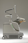 From Philips Medical Systems;
'The Philips iU22 system combines Intelligent Design, including breakthroughs in ergonomics, with Intelligent Control, providing new levels of automation, to give you revolutionary performance and workflow.'
Device Information and Specification
APPLICATIONS
Abdominal, cardiac (also for adults with TEE), musculoskeletal (also pediatric), OB/GYN, prostate, smallparts, transcranial, vascular
CONFIGURATION
17' high resolution non-interlaced flat CRT, 4 active probe ports
B-mode, M-mode, coded harmonic imaging, color flow mode (CFM), power Doppler imaging (PDI), color Doppler, pulsed wave Doppler, tissue harmonic imaging
IMAGING OPTIONS
CrossXBeam spatial compounding, coded ultrasound acquisition),speckle reduction imaging (SRI), TruScan technology store raw data, CINE review with 4 speed types
OPTIONAL PACKAGE
Transesophageal scanning, stress echo, tissue velocity imaging (TVI), tissue velocity Doppler (TVD), contrast harmonic imaging
STORAGE, CONNECTIVITY, OS
Patient and image archive, HDD, DICOM 3.0, CD/DVD, MOD, Windows-based
DATA PROCESSING
Digital beamformer with 1024 system processing channel technology
H*W*D m (inch.)
1.62 * 0.61 * 0.99 (64 * 22 * 43)
WEIGHT
kg (345 lbs.)
POWER CONSUMPTION
Result Pages : | Share This Page Look Ups |
Medical-Ultrasound-Imaging.com
former US-TIP.com
Member of SoftWays' Medical Imaging Group - MR-TIP • Radiology TIP • Medical-Ultrasound-Imaging
Copyright © 2008 - 2024 SoftWays. All rights reserved.
Terms of Use | Privacy Policy | Advertise With Us
former US-TIP.com
Member of SoftWays' Medical Imaging Group - MR-TIP • Radiology TIP • Medical-Ultrasound-Imaging
Copyright © 2008 - 2024 SoftWays. All rights reserved.
Terms of Use | Privacy Policy | Advertise With Us
[last update: 2023-11-06 01:42:00]