Medical Ultrasound Imaging
Sunday, 19 May 2024
Info Sheets Out- side     | 'Pregnancy Ultrasound' p2 Searchterm 'Pregnancy Ultrasound' found in 17 articles 1 term [ • ] - 6 definitions [• ] - 10 booleans [• ]Result Pages : •
As far as ultrasound is concerned, 4D ultrasound (also referred to as live 3D ultrasound or 4B-mode) is the latest ultrasound technology - the fourth dimension means length, width, and depth over time. 4D Ultrasound takes 3D ultrasound images and adds the element of time to the progress so that a moving three-dimensional image is seen on the monitor. A 4D scan takes the same amounts of time as a 2D or 3D scan; the difference is the ultrasound equipment being used. One advantage of a 4D fetal ultrasound to a 2D-mode is that parents can see how their baby will generally look like. However, there are different opinions over the medical advantages. To scan a 3D ultrasound image, the probe is swept over the maternal abdomen. A computer takes multiple images and renders the 3D picture. With 4D imaging, the computer takes the images as multiple pictures while the probe is hold still and a 3D image is simultaneously rendered in real time on a monitor. In most cases, the standard 2D ultrasound is taken, and then the 3D/4D scan capability is added if an abnormality is detected or suspected. The 3D/4D sonogram is then focused on a specific area, to provide the details needed to assess and diagnose a suspected problem. A quick 4D scan of the face of the fetus may be performed at the end of a routine exam, providing the parents with a photo. See also Obstetric and Gynecologic Ultrasound, Pregnancy Ultrasound, Fetal Ultrasound and Abdominal Ultrasound. •
(US) Also called echography, sonography, ultrasonography, echotomography, ultrasonic tomography. Diagnostic imaging plays a vital role in modern healthcare, allowing medical professionals to visualize internal structures of the body and assist in the diagnosis and treatment of various conditions. Two terms that are commonly used interchangeably but possess distinct meanings in the field of medical imaging are 'ultrasound' and 'sonography.' Ultrasound is the imaging technique that utilizes sound waves to create real-time images, while sonography encompasses the entire process of performing ultrasound examinations and interpreting the obtained images. Ultrasonography is a synonymous term for sonography, emphasizing the use of ultrasound technology in diagnostic imaging. A sonogram, on the other hand, refers to the resulting image produced during an ultrasound examination. Ultrasonic waves, generated by a quartz crystal, cause mechanical perturbation of an elastic medium, resulting in rarefaction and compression of the medium particles. These waves are reflected at the interfaces between different tissues due to differences in their mechanical properties. The transmission and reflection of these high-frequency waves are displayed with different types of ultrasound modes. By utilizing the speed of wave propagation in tissues, the time of reflection information can be converted into distance of reflection information. The use of higher frequencies in medical ultrasound imaging yields better image resolution. However, higher frequencies also lead to increased absorption of the sound beam by the medium, limiting its penetration depth. For instance, higher frequencies (e.g., 7.5 MHz) are employed to provide detailed imaging of superficial organs like the thyroid gland and breast, while lower frequencies (e.g., 3.5 MHz) are used for abdominal examinations. Ultrasound in medical imaging offers several advantages including:
•
noninvasiveness;
•
safety with no potential risks;
•
widespread availability and relatively low cost.
Diagnostic ultrasound imaging is generally considered safe, with no adverse effects. As medical ultrasound is extensively used in pregnancy and pediatric imaging, it is crucial for practitioners to ensure its safe usage. Ultrasound can cause mechanical and thermal effects in tissue, which are amplified with increased output power. Consequently, guidelines for the safe use of ultrasound have been issued to address the growing use of color flow imaging, pulsed spectral Doppler, and higher demands on B-mode imaging. Furthermore, recent ultrasound safety regulations have shifted more responsibility to the operator to ensure the safe use of ultrasound. See also Skinline, Pregnancy Ultrasound, Obstetric and Gynecologic Ultrasound, Musculoskeletal and Joint Ultrasound, Ultrasound Elastography and Prostate Ultrasound. Further Reading: Basics: News & More:
• 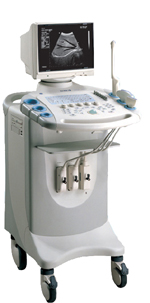
From SIUI Inc.;
Device Information and Specification
CONFIGURATION
Normal system, gray scale(256)
Linear and convex
PROBES STANDARD
2.5MHz ~ 11.0MHz, broad band, tri-frequency
IMAGING OPTIONS
Real zoom, max. Zoom * 4.0, multiplying power and position selectable
OPTIONAL PACKAGE
H*W*D m
1.30 * 0.52 * 0.77
WEIGHT
90kg (main unit)
POWER REQUIREMENT
AC 220V/110V, 50Hz/60Hz
POWER CONSUMPTION
0.45 KVA
•  From SIUI Inc.;
'Dedicated to ultrasound industry, Shantou Institute of Ultrasonic Instruments, Inc. (SIUI) has launched Apogee 3500, the Digital Color Doppler Ultrasound Imaging System. With latest imaging technologies, high-definition image quality and excellent practical functions, the Apogee 3500 offers optimal solutions for clinical ultrasonic examination.' 'The Apogee 3500 is available with many high-density, super broadband and multi-frequency probes, such as convex, micro-convex, linear, vaginal, rectal and phased array probes, which are widely applied for different clinical diagnoses, including abdomen (liver, kidney, gall-bladder, pancreas), gynecology (uterus, ovary), obstetrics (early pregnancy, basic OB, complete OB, multi gestation, fetal echo), cardiology (adult and pediatric cardiology), small parts (thyroid, galactophore, testicles, neonate), peripheral vascular and prostate.'
Device Information and Specification
CONFIGURATION
Normal system, color - gray scale(256)
Linear, convex and phased array
PROBES STANDARD
2.0 MHz ~ 12.0 MHz, broad band, tri-frequency
B-mode (B, 2B, 4B), M-mode, B/M-mode, real-time compound imaging, panoramic imaging, trapezoidal imaging (linear probes), spectrum Doppler (PWD and CWD), color Doppler flow imaging (CDFI), color power angio (CPA), tissue harmonic imaging (THI)
IMAGING OPTIONS
Real-time ZOOM, zoom rate and position selectable
OPTIONAL PACKAGE
H*W*D m
1.29 * 0.52 * 0.75
WEIGHT
110 kg
POWER REQUIREMENT
AC 220V/110V, 50Hz/60Hz
POWER CONSUMPTION
0.6 KVA
•
A-mode (Amplitude-mode) ultrasound is a technique used to assess organ dimensions and determine the depth of an organ. While A-mode technology was previously employed in midline echoencephalography for rapid screening of intracranial mass lesions and ophthalmologic scanning, it is now considered obsolete in medical imaging. Nonetheless, the A-mode scan has found applications in early pregnancy assessment (specifically the detection of fetal heartbeats), cephalometry, and placental localization.
When the ultrasound beam encounters an anatomic boundary, the received sound impulse is processed to appear as a vertical reflection of a point. On the display, it looks like spikes of different heights (the amplitude). The intensity of the returning impulse determined the height of the vertical reflection and the time it took for the impulse to make the round trip would determine the space between verticals. The distance between these spikes can be measured accurately by dividing the speed of sound in tissue (1540 m/sec) by half the sound travel time. During an echoencephalography scan, the first A-mode scan is acquired from the right side of the head and captured on film. Subsequently, the probe is positioned at the corresponding point on the left side, and a second exposure is captured on the same film, displaying inverted spikes. The A-mode ultrasound could be used to identify structures normally located in the midline of the brain such as the third ventricle and falx cerebri. The midline structures would be aligned in normal patients but show displacement in patients with mass lesion such as a subdural, epidural, or intracranial hemorrhage. See also 2D Ultrasound, 3D Ultrasound, 4D Ultrasound, Ultrasound Biomicroscopy, A-scan, B-mode and the Infosheet about ultrasound modes. Result Pages : | Share This Page Look Ups |
Medical-Ultrasound-Imaging.com
former US-TIP.com
Member of SoftWays' Medical Imaging Group - MR-TIP • Radiology TIP • Medical-Ultrasound-Imaging
Copyright © 2008 - 2024 SoftWays. All rights reserved.
Terms of Use | Privacy Policy | Advertise With Us
former US-TIP.com
Member of SoftWays' Medical Imaging Group - MR-TIP • Radiology TIP • Medical-Ultrasound-Imaging
Copyright © 2008 - 2024 SoftWays. All rights reserved.
Terms of Use | Privacy Policy | Advertise With Us
[last update: 2023-11-06 01:42:00]