Medical Ultrasound Imaging
Sunday, 19 May 2024
Info Sheets Out- side     | 'Range' p4 Searchterm 'Range' found in 102 articles 4 terms [ • ] - 98 definitions [• ] Result Pages : •
To calculate the echo position, a constant sound speed of 1538.5 m/sec is assumed. Tissue penetration is frequency depended, if the frequency increases, the imaging depth decreases. The range resolution defines the depth. Ultrasound propagating in tissue is attenuated due to scattering and absorption. The attenuation is proportional to depth and frequency and is typically in the range from 0.5 to 1 dB/(MHz cm). See also Attenuation Coefficient, Proximity Sensor, and Echo Ranging. •
Doppler techniques are dependent on the transducers used. The transducer operating in continuous wave mode utilizes one half of the elements and is continuously sending sound energy while the other half is continuously receiving the reflected signals. If the transducer is being used in a pulsed wave mode, the whole transducer is used to send and then receive the returning signals. Pulsed wave techniques allow the accurate measurement of blood flow at a specific area in the heart and the detection of both velocity and direction. Measurement is performed by timing the reception of the returning signals giving a view of flows at specific depths. The region where flow velocities are measured is called the sample volume. Errors in the accuracy of the information arise if the velocities exceed a certain speed. The highest velocity accurately measured is called the Nyquist limit.
•
Continuous Wave Doppler
Used for accurate measurement of high Velocity flow. A disadvantage is the poor range of resolution.
•
Pulsed Wave Doppler
Used for the measurement of velocities at a specific location with a good range of resolution. A disadvantage is the imprecise measuring of high velocities. See also Doppler Velocity Signal and Doppler Effect. Further Reading: Basics:
News & More:
• 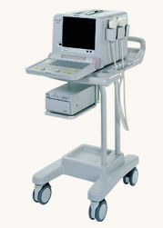 From Hitachi Medical Systems America Inc.;
The EUB-405 PLUS ultrasound system is a small portable unit and offers extreme dependability and uncompromising image quality. Compatible with a wide range of transducers, this system is a cost effective solution for a variety of clinical settings. A variety of measurements and calculations including distance, area, circumference, volume, are possible. Device Information and Specification
APPLICATIONS
General purpose, OB, urological
CONFIGURATION
Compact portable system
Dual frequency
Linear, convex, radial, bi-plane, echoendoscope longitudinal
PROBE PORTS
One
OPTIONAL PACKAGE
Cine/multi-memory, digital image storage and transfer via flashcard, cart, second port
WEIGHT
Less than 14 kg
• 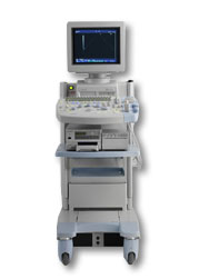 From Hitachi Medical Corporation (HMC), sales, marketing and service in the US by Hitachi Medical Systems America Inc.
The HI VISION™ 5500 - EUB-5500 fully digital ultrasound system delivers the latest generation of signal processing technology, sophisticated transducer design, and a host of features and options for advanced imaging capabilities across a wide range of clinical situations. This system is compatible with all Pentax ultrasound endoscopes.
Device Information and Specification
APPLICATIONS
Abdominal, brachytherapy/cryotherapy, breast, cardiac, dedicated biopsy, endoscopic, intraoperative, laparoscopic, musculoskeletal, OB/GYN, pediatric, small parts, urologic, vascular
CONFIGURATION
Compact system
Five frequency (except mini-probes)
Linear, convex, radial, miniradial/miniprobe, biplane, phased array, echoendoscope longitudinal, echoendoscope radial
3 modes of dynamic tissue harmonic imaging (dTHI), pulsed wave Doppler, continuous wave Doppler, color flow imaging, power Doppler, directional power Doppler, color flow angiography, real-time Doppler measurements
IMAGING OPTIONS
3RD generation color artifact suppression
OPTIONAL PACKAGE
STORAGE, CONNECTIVITY, OS
Patient and image database management system, HDD, FDD, MOD, CD-ROM, Network, DICOM 3.0, Windows XP
DATA PROCESSING
H*W*D m (inch.)
1.40 x 0.51 x 0.79 (55 x 20 x 31)
WEIGHT
130 kg (286 lbs.)
POWER CONSUMPTION
1.2kVA
ENVIRONMENTAL IMPACT
4096 btu/hr heat output
•
The earliest introduction of vascular ultrasound contrast agents (USCA) was by Gramiak and Shah in 1968, when they injected agitated saline into the ascending aorta and cardiac chambers during echocardiographic to opacify the left heart chamber. Strong echoes were produced within the heart, due to the acoustic mismatch between free air microbubbles in the saline and the surrounding blood. The disadvantage of this microbubbles produced by agitation, was that the air quickly leak from the thin bubble shell into the blood, where it dissolved. In addition, the small bubbles that were capable of traversing the capillary bed did not survive long enough for imaging because the air quickly dissipated into the blood. Aside from agitated saline, also hydrogen peroxide, indocyanine green dye, and iodinated contrast has been tested. The commercial development of contrast agents began in the 1980s with greatest effort to the stabilization of small microbubbles. The development generations by now:
•
first generation USCA = non-transpulmonary vascular;;
•
second generation USCA = transpulmonary vascular, with short half-life (less than 5 min);
•
third generation USCA = transpulmonary vascular, with longer half-life (greater than 5 min).
To pass through the lung capillaries and enter into the systemic circulation, microspheres should be less than 10 μm in diameter. Air bubbles in that size range persist in solution for only a short time; too short for systemic vascular use. The first developed agent was Echovist (1982), which enabled the enhancement of the right heart. The second generation of echogenic agents, sonicated 5% human albumin-containing air bubbles (Albunex), were capable of transpulmonary passage but often failed to produce adequate imaging of the left heart. Both Albunex and Levovist utilize air as the gas component of the microbubble. In the 1990s newer developed agents with fluorocarbon gases and albumin, surfactant, lipid, or polymer shells have an increased persistence of the microspheres. This smaller, more stable microbubble agents, and improvements in ultrasound technology, have resulted in a wider range of application including myocardial perfusion. See also First Generation USCA, Second Generation USCA, and Third Generation USCA. Further Reading: Basics:
Result Pages : | Share This Page Look Ups |
Medical-Ultrasound-Imaging.com
former US-TIP.com
Member of SoftWays' Medical Imaging Group - MR-TIP • Radiology TIP • Medical-Ultrasound-Imaging
Copyright © 2008 - 2024 SoftWays. All rights reserved.
Terms of Use | Privacy Policy | Advertise With Us
former US-TIP.com
Member of SoftWays' Medical Imaging Group - MR-TIP • Radiology TIP • Medical-Ultrasound-Imaging
Copyright © 2008 - 2024 SoftWays. All rights reserved.
Terms of Use | Privacy Policy | Advertise With Us
[last update: 2023-11-06 01:42:00]