Medical Ultrasound Imaging
Sunday, 19 May 2024
Info Sheets Out- side     | 'Sampling Rate' p2 Searchterm 'Sampling Rate' found in 9 articles 1 term [ • ] - 3 definitions [• ] - 5 booleans [• ]Result Pages : •
The M-mode (Motion-mode) ultrasound is used for analyzing moving body parts (also called time-motion or TM-mode) commonly in cardiac and fetal cardiac imaging. The application of B-mode and a strip chart recorder allows visualization of the structures as a function of depth and time. The M-mode ultrasound transducer beam is stationary while the echoes from a moving reflector are received at varying times.
A single beam in an ultrasound scan is used to produce the one-dimensional M-mode picture, where movement of a structure such as a heart valve can be depicted in a wave-like manner. The high sampling frequency (up to 1000 pulses per second) is useful in assessing rates and motion, particularly in cardiac structures such as the various valves and the chamber walls. Further Reading: News & More:
• 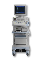 From Hitachi Medical Corporation (HMC), sales, marketing and service in the US by Hitachi Medical Systems America Inc.
The HI VISION™ 5500 - EUB-5500 fully digital ultrasound system delivers the latest generation of signal processing technology, sophisticated transducer design, and a host of features and options for advanced imaging capabilities across a wide range of clinical situations. This system is compatible with all Pentax ultrasound endoscopes.
Device Information and Specification
APPLICATIONS
Abdominal, brachytherapy/cryotherapy, breast, cardiac, dedicated biopsy, endoscopic, intraoperative, laparoscopic, musculoskeletal, OB/GYN, pediatric, small parts, urologic, vascular
CONFIGURATION
Compact system
Five frequency (except mini-probes)
Linear, convex, radial, miniradial/miniprobe, biplane, phased array, echoendoscope longitudinal, echoendoscope radial
3 modes of dynamic tissue harmonic imaging (dTHI), pulsed wave Doppler, continuous wave Doppler, color flow imaging, power Doppler, directional power Doppler, color flow angiography, real-time Doppler measurements
IMAGING OPTIONS
3RD generation color artifact suppression
OPTIONAL PACKAGE
STORAGE, CONNECTIVITY, OS
Patient and image database management system, HDD, FDD, MOD, CD-ROM, Network, DICOM 3.0, Windows XP
DATA PROCESSING
H*W*D m (inch.)
1.40 x 0.51 x 0.79 (55 x 20 x 31)
WEIGHT
130 kg (286 lbs.)
POWER CONSUMPTION
1.2kVA
ENVIRONMENTAL IMPACT
4096 btu/hr heat output
•  From Hitachi Medical Corporation (HMC);
The HI VISION™ 6500 - EUB-6500 high resolution digital ultrasound system offers advanced clinical imaging, enhanced operating efficiency, and remarkable clinical flexibility, all in robust and versatile configuration that simply represents a better clinical solution in a variety of real-world, real-work arenas.
Device Information and Specification
APPLICATIONS
Abdominal, brachytherapy/cryotherapy, breast, cardiac, dedicated biopsy, endoscopic, intraoperative, laparoscopic, musculoskeletal, OB/GYN, pediatric, small parts, urologic, vascular
CONFIGURATION
Compact system
Linear, convex, radial, miniradial/miniprobe, biplane, phased array, echoendoscope longitudinal, echoendoscope radial
Tissue Doppler imaging (TDI), pulsed wave Doppler, continuous wave Doppler, color flow imaging, power Doppler, directional power Doppler, color flow angiography, real-time Doppler measurements, 4 modes of dynamic tissue harmonic imaging (dTHI), wideband pulse inversion imaging (WPI)
IMAGING OPTIONS
3RD generation color artifact suppression
OPTIONAL PACKAGE
3D ultrasound, dual omni-directional M-mode display, steerable CW Doppler, dynamic contrast harmonics imaging, stress echo, Pentax EUS and Fujinon Mini-probe
STORAGE, CONNECTIVITY, OS
Patient and image database management system, HDD, FDD, MOD, CD-ROM, Network, DICOM 3.0, Windows XP
DATA PROCESSING
H*W*D m (inch.)
1.40 x 0.51 x 0.79 (55 x 20 x 31)
WEIGHT
130 kg (286 lbs.)
POWER CONSUMPTION
1.2kVA
ENVIRONMENTAL POLLUTION
4096 btu/hr heat output
•
Pregnancy ultrasound plays a crucial role in monitoring the health and development of the fetus throughout pregnancy. It serves as a screening tool with various applications, including:
•
Verification of Due Date and Assessment of Pregnancy Health: Fetal ultrasound examinations are used to accurately determine the estimated due date of the baby. They also aid in investigating the causes of bleeding during pregnancy and assessing the overall health and well-being of the fetus. •
•
Measurement of Amniotic Fluid and Placental Assessment: Ultrasound is utilized to measure the amniotic fluid levels, which provide insights into fetal well-being and the functioning of the placenta. It also helps evaluate the condition of the placenta, ensuring proper nutrient and oxygen supply to the developing baby. •
Early Pregnancy Confirmation and Multiple Fetuses Detection: Around week five to seven of pregnancy, ultrasound is utilized to confirm the pregnancy, determine the fetal size, and detect the presence of multiple fetuses. It aids in distinguishing between intrauterine and ectopic pregnancies, ensuring appropriate management. •
Third-Trimester Evaluation: As the pregnancy progresses, ultrasound assessments are conducted to evaluate fetal size, position, growth, and the condition of the placenta. This information assists healthcare providers in monitoring the well-being of the fetus and planning for a safe delivery. •
Guiding Procedures: Ultrasound plays a vital role in guiding invasive procedures such as amniocentesis and chorionic villus sampling. It helps guide the placement of a needle to collect cells from the amniotic fluid or placenta, aiding in genetic testing and diagnosing potential fetal abnormalities. See also Doppler Fluximetry in Pregnancy, Fetal Ultrasound, Obstetric and Gynecologic Ultrasound and Vaginal Probe. Result Pages : | Share This Page Look Ups |
Medical-Ultrasound-Imaging.com
former US-TIP.com
Member of SoftWays' Medical Imaging Group - MR-TIP • Radiology TIP • Medical-Ultrasound-Imaging
Copyright © 2008 - 2024 SoftWays. All rights reserved.
Terms of Use | Privacy Policy | Advertise With Us
former US-TIP.com
Member of SoftWays' Medical Imaging Group - MR-TIP • Radiology TIP • Medical-Ultrasound-Imaging
Copyright © 2008 - 2024 SoftWays. All rights reserved.
Terms of Use | Privacy Policy | Advertise With Us
[last update: 2023-11-06 01:42:00]