Medical Ultrasound Imaging
Saturday, 18 May 2024
Info Sheets Out- side     | 'Spectra' p4 Searchterm 'Spectra' found in 30 articles 4 terms [ • ] - 26 definitions [• ] Result Pages : •
Duplex ultrasonography (duplex scan) consists of two ultrasound modalities to study blood flow and the perivascular tissue. This includes B-mode / gray scale imaging used in combination with spectral Doppler / pulsed-wave Doppler. The real-time visualization of the vessels and tissue by the B-mode component improves the PW Doppler positioning and the direction of blood flow can be inferred. The angle between the direction of the PW Doppler signal and the estimated direction of blood flow can be measured. Duplex techniques are available on phased array, linear array, and mechanical scanners. A phased array probe is able to create nearly simultaneous images and flow information. A linear array transducer can also do this if the Doppler probe is attached separately to one end of the scanhead. A mechanical transducer freeze the image; the crystals must be static to produce a Doppler image. The first two transducers are therefore the best choice for Duplex. See also Compound B-Mode, and Duplex Scanner. Further Reading: News & More:
•
Duplex systems often combine a pulsed Doppler with a spectral display and a real time imaging system. A duplex scanner has usually an imaging transducer or a separate transducer used to collect continuous wave or pulsed Doppler signals, either simultaneously with imaging or sequentially. •
Harmonic imaging relies on detection of harmonics of the transmitted
frequency produced by bubble oscillation. This method is widely available on ultrasound scanners and uses the same array transducers as conventional imaging. A major limitation of the use of ultrasound contrast agents is the problem that signals from the microbubbles are mixed with those from tissue. Echoes from solid tissue and red blood cells are suppressed by harmonic imaging. In harmonic mode, the system transmits at one frequency, but is tuned to receive echoes preferentially at double that frequency, and the second harmonic echoes from the place of the bubble. Typically, the transmit frequency lies between 1.5 and 3 MHz and the receive frequency is selected by means of a bandpass filter whose center frequency lies between 3 and 6 MHz. Color Doppler and real-time harmonic spectral Doppler modes have also been implemented and show a level of tissue motion suppression not available in conventional modes. See also Harmonic B-Mode Imaging, and Harmonic Power Doppler. Further Reading: Basics:
News & More:
• 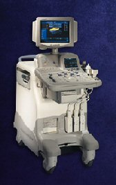 From GE Healthcare.;
Versatile High Performance System 'The LOGIQ 5 Expert ultrasound system delivers the premium performance advantage of TruScan architecture in a versatile high performance package. Advanced capabilities for outstanding clinical performance are available with the efficiency and productivity needed to meet clinical demands.'
Device Information and Specification
APPLICATIONS
Abdominal, cardiac, musculoskeletal, neonatal, OB/GYN, small parts, transcranial, urologic, vascular
CONFIGURATION
Normal system
B-mode, M-mode, anatomic M-mode creation and adjustment, triplex, 3D ultrasound, pulsed wave Doppler, continuous wave Doppler, power Doppler, color Doppler, spectral Doppler
IMAGING OPTIONS
OPTIONAL PACKAGE
STORAGE, CONNECTIVITY, OS
On-board patient, image and reporting archive, HDD, CD-ROM disk burner included, PCMCIA, USB, Windows-based OS
DATA PROCESSING
H*W*D m (inch.)
1.45 * 0.52 * 0.99 (57 * 20 * 39)
WEIGHT
180 kg (397 lbs.)
POWER CONSUMPTION
less than 1.5 KVA
• Result Pages : | Share This Page Look Ups |
Medical-Ultrasound-Imaging.com
former US-TIP.com
Member of SoftWays' Medical Imaging Group - MR-TIP • Radiology TIP • Medical-Ultrasound-Imaging
Copyright © 2008 - 2024 SoftWays. All rights reserved.
Terms of Use | Privacy Policy | Advertise With Us
former US-TIP.com
Member of SoftWays' Medical Imaging Group - MR-TIP • Radiology TIP • Medical-Ultrasound-Imaging
Copyright © 2008 - 2024 SoftWays. All rights reserved.
Terms of Use | Privacy Policy | Advertise With Us
[last update: 2023-11-06 01:42:00]