Medical Ultrasound Imaging
Sunday, 19 May 2024
Info Sheets Out- side     | 'Transducer Types' p3 Searchterm 'Transducer Types' found in 17 articles 1 term [ • ] - 2 definitions [• ] - 14 booleans [• ]Result Pages : • 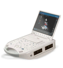 From ESAOTE S.p.A.;
'The MyLab™30CV ultrasound system is an evolutionary step in ultrasound technology. Weighing less than 20 pounds, it is the first compact ultrasound system to deliver premium console performance. And with mobile, portable or stationary configurations, MyLab30CV can adapt to any clinical environment.'
Device Information and Specification
APPLICATIONS
Abdominal, breast, cardiac, OB/GYN, pediatric, pediatric cardiology, small parts, transcranial, vascular
CONFIGURATION
Portable
Linear: 4-10 MHz, convex: 2-5 MHz, phased: 1.6-10 MHz, micro convex: 5-7.5 MHz, endocavity: 5-7.5 MHz, pencil: 2 + 5 MHz
2-D, M-mode, duplex, triplex, color Doppler, pulsed wave Doppler, tissue velocity mapping (TVM), tissue enhancement imaging (TEI™), contrast harmonic imaging, stress echo, tissue velocity mapping for LV motion analysis (TVM), contrast tuned imaging for contrast media procedures (CnTI™), Qontrast™ for myocardium parameters quantification
STORAGE, CONNECTIVITY, OS
Digital patient archive/management, integrated CD/RW, RJ 45 and USB ports, Windows
H*W*D m (inch.)
0.16 * 0.36 * 0.50 (6.2 x 14 x 19.3)
WEIGHT
Less than 11 kg (20 lbs.)
• 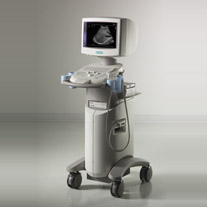 From Siemens Medical Systems;
'The SONOLINE G20™ ultrasound system quickly distances itself from the competition with next-generation all-digital system architecture that utilizes Siemens technology migration. Individual imaging parameters have been optimized for a wide variety of clinical applications and patient types. So you can realize a higher degree of diagnostic confidence. Without doubt.'
Device Information and Specification
CONFIGURATION
Compact, ultra-portable system
MultiHertz™ multiple frequency
PROBE TYPES
MicroCase™ transducer
IMAGING OPTIONS
Tissue Harmonic Imaging (THI)
IMAGING ENHANCEMENTS
TGO™ tissue grayscale optimization technology
STORAGE
DIMAQ-IP integrated workstation
DATA PROCESSING
Powerful processor for rapid transition times
• 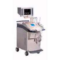 From Siemens Medical Systems;
'The SONOLINE Omnia™ ultrasound system offers mobility, high performance, and ease of use. This digital imaging system delivers excellent 2D, color flow, and Doppler image quality for a variety of general imaging exam types. In addition, the SONOLINE Omnia with cardiac option includes special features that enable use for adult cardiac imaging.'
Device Information and Specification
CLINICAL APPLICATION
Abdomen, small parts, pediatric, prostate, orthopedic, obstetrics, gynecology, cerebrovascular, musculoskeletal, rectal, peripheral vascular (venous and arterial), cardiology
CONFIGURATION
Compact, mobile system
Multi-Frequency and wideband
Wide range of linear/curved/phased array, mechanical, CW pencil probes, laparoscopic, intraoperative, biopsy, TEE transducers
PROBE PORTS
Five
IMAGING OPTIONS
OPTIONAL PACKAGE
Upgradeable applications, cardiac option
IMAGING ENHANCEMENTS
Ultra Fast 3D rendering
STORAGE
Magneto-Optical Drive of 640 MB
DATA PROCESSING
MultiDimensional image processor
•
Ultrasound elastography is a specialized imaging technique that provides information about tissue elasticity or stiffness. It is used to assess the mechanical properties of tissues, helping to differentiate between normal and abnormal tissue conditions.
The basic principle behind ultrasound elastography involves the application of mechanical stress to the tissue and measuring its resulting deformation. This is typically achieved by using either external compression or shear waves generated by the ultrasound transducer. There are two main types of ultrasound elastography: •
Strain Elastography: In strain elastography, the tissue is mechanically compressed using the ultrasound transducer, causing deformation. The transducer then captures images before and after compression, and the software analyzes the displacement or strain between these images. Softer tissues tend to deform more than stiffer tissues, and this information is used to generate a color-coded map or elastogram, where softer areas appear in different colors compared to stiffer regions.
•
Shear Wave Elastography: Shear wave elastography involves the generation of shear waves within the tissue using focused ultrasound beams. These shear waves propagate through the tissue, and their velocity is measured using the ultrasound transducer. The speed of shear wave propagation is directly related to tissue stiffness: stiffer tissues transmit shear waves faster than softer tissues. By calculating the shear wave velocity, an elastogram is generated, providing a quantitative assessment of tissue stiffness.
Both strain elastography and shear wave elastography offer valuable insights into tissue characteristics and can assist in the diagnosis and characterization of various conditions. In clinical practice, ultrasound elastography is particularly useful for evaluating liver fibrosis, breast lesions, thyroid nodules, prostate abnormalities, and musculoskeletal conditions. By providing additional information about tissue stiffness, ultrasound elastography enhances the diagnostic capabilities of traditional ultrasound imaging. It allows for non-invasive assessment, improves the accuracy of tissue characterization, and aids in treatment planning and monitoring of various medical conditions. See also Ultrasound Accessories and Supplies, Sonographer and Ultrasound Technology. •
Ultrasound machines, widely used in medical imaging, are essential tools in the field of diagnostic ultrasound. These devices utilize high-frequency sound waves to create real-time images of internal body structures. Ultrasound machines consist of several key components that work together to generate diagnostic images.
These include:
•
The transducer is a handheld device that emits and receives sound waves. It converts electrical energy into sound waves and captures the returning echoes to create images.
•
The control panel houses the interface where the sonographer adjusts imaging parameters such as depth, frequency, and gain. It allows for customization of imaging settings based on the clinical requirements. The transducer pulse controls change the amplitude, frequency and duration of the pulses emitted from the transducer probe.
•
The central processing unit (CPU) serves as the brain of the ultrasound machine, processing the acquired data and transforming it into images. It handles complex calculations, image optimization, data storage and contains the electrical power supplies for itself and the transducer probe.
•
The display monitor (oscilloscope, tablet, computer monitor, etc.) showcases the real-time ultrasound images produced by the machine. It provides visual feedback to the sonographer, aiding in the interpretation and analysis of anatomical structures. Handheld ultrasound devices and mobile ultrasound probes can be connected wirelessly to a smartphone or tablet via Bluetooth or WiFi. These end device serves then as the ultrasound monitor.
•
Data input and measurements are done with the keyboard cursor (trackball). Ultrasound devices used for handheld point of care ultrasound (HPOCUS) are operated via the touch screen of the control panel.
•
Images are captured, reviewed, stored and transmitted digitally, using a standard format for digital imaging and communications in medicine (DICOM). Disk storage devices (FDD, HDD, CD, DVD) are outdated, but may be used in older machines to store the acquired images if no picture archiving and communication system (PACS) connection is possible.
•
The displayed ultrasound pictures are usually digitally stored in a PACS. The images from portable ultrasound machines can be stored and conveniently managed on the end device itself, the inserted memory card or in the cloud. With a QR scanner, the images can be accessed via the Internet in the cloud. Often there is also the possibility to get a picture of a baby sonography as a printout.
B-mode machines represent the vast majority of machines used in echocardiology, obstetrical scans, abdominal scans, gynecological scans, etc. B-mode ultrasound machines usually produce the sector (or pie segment-shaped) scans. These ultrasound scans require either a mechanical scanner transducer (the transducer moves to produce the sector scan), or a linear array transducer operated as a phased array. Ultrasound machines come in different types, each catering to specific clinical needs. The two primary types are stationary and portable ultrasound machines: •
Stationary units are typically larger in size and are installed in dedicated imaging rooms. These machines offer advanced imaging capabilities and a wide range of specialized features. They are commonly found in hospitals, clinics, and university medical centers where comprehensive imaging services are provided.
•
Portable units (see Portable Ultrasound Machine), as the name suggests, are compact and lightweight, designed for on-the-go imaging. These machines are highly versatile and offer excellent mobility, allowing healthcare professionals to bring the ultrasound system directly to the patient's bedside. Portable ultrasound machines are particularly useful in emergency settings, rural healthcare facilities, and point-of-care applications.
See also Handheld Ultrasound, Ultrasound System Performance, Equipment Preparation, Coaxial Cable, and Microbubble Scanner Modification, Environmental Protection and Ultrasound Accessories and Supplies. Further Reading: Basics: News & More:
Result Pages : | Share This Page Look Ups |
Medical-Ultrasound-Imaging.com
former US-TIP.com
Member of SoftWays' Medical Imaging Group - MR-TIP • Radiology TIP • Medical-Ultrasound-Imaging
Copyright © 2008 - 2024 SoftWays. All rights reserved.
Terms of Use | Privacy Policy | Advertise With Us
former US-TIP.com
Member of SoftWays' Medical Imaging Group - MR-TIP • Radiology TIP • Medical-Ultrasound-Imaging
Copyright © 2008 - 2024 SoftWays. All rights reserved.
Terms of Use | Privacy Policy | Advertise With Us
[last update: 2023-11-06 01:42:00]