Medical Ultrasound Imaging
Sunday, 19 May 2024
Info Sheets Out- side     | 'Ultrasound' p17 Searchterm 'Ultrasound' found in 466 articles 60 terms [ • ] - 406 definitions [• ] Result Pages : •
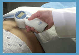
A transducer is a device, usually electrical or electronic, that converts one type of energy to another. Most transducers are either sensors or actuators. A transducer (also called probe) is a main part of the ultrasound machine. The transducer sends ultrasound waves into the body and receives the echoes produced by the waves when it is placed on or over the body part being imaged. Ultrasound transducers are made from crystals with piezoelectric properties. This material vibrates at a resonant frequency, when an alternating electric current is applied. The vibration is transmitted into the tissue in short bursts. The speed of transmission within most soft tissues is 1540 m/s, producing a transit time of 6.5 ms/cm. Because the velocity of ultrasound waves is constant, the time taken for the wave to return to the transducer can be used to determine the depth of the object causing the reflection. The waves will be reflected when they encounter a boundary between two tissues of different density (e.g. soft tissue and bone) and return to the transducer. Conversely, the crystals emit electrical currents when sound or pressure waves hit them (piezoelectric effect). The same crystals can be used to send and receive sound waves; the probe then acts as a receiver, converting mechanical energy back into an electric signal which is used to display an image. A sound absorbing substance eliminates back reflections from the probe itself, and an acoustic lens focuses the emitted sound waves. Then, the received signal gets processed by software to an image which is displayed at a monitor. Transducer heads may contain one or more crystal elements. In multi-element probes, each crystal has its own circuit. The advantage is that the ultrasound beam can be controlled by changing the timing in which each element gets pulsed. Especially for cardiac ultrasound it is important to steer the beam. Usually, several different transducer types are available to select the appropriate one for optimal imaging. Probes are formed in many shapes and sizes. The shape of the probe determines its field of view. Transducers are described in megahertz (MHz) indicating their sound wave frequency. The frequency of emitted sound waves determines how deep the sound beam penetrates and the resolution of the image. Most transducers are only able to emit one frequency because the piezoelectric ceramic or crystals within it have a certain inherent frequency, but multi-frequency probes are also available. See also Blanking Distance, Damping, Maximum Response Axis, Omnidirectional, and Huygens Principle. Further Reading: News & More:
•
Attenuation is the reduction of power, for example due to the passage through a medium or electrical component. In ultrasound imaging, attenuation means the decrease in amplitude and intensity as a sound wave travels through a medium. In ultrasound attenuation is often characterized as the half-value layer, or the half-power distance. These terms refer to the distance that ultrasound will travel in a particular tissue before its energy is attenuated to half its original value. Attenuation originates through:
•
divergence of the wavefront;
•
absorption of wave energy;
•
elastic reflection of wave energy;
•
elastic scattering of wave energy.
A thick muscled chest wall will offer a significant obstacle to the transmission of ultrasound. Non-muscle tissue such as fat does not attenuate acoustic energy as much. The half-value layer for bone is still less than muscle, that's why bone is such a barrier to ultrasound. See also Attenuation Coefficient, and Derated Quantity. Further Reading: Basics:
•
Also called B-mode echography, B-mode sonography, 2D-mode, and sonogram. B-mode ultrasound (Brightness-mode) is the display of a 2D-map of B-mode data, currently the most common form of ultrasound imaging. The development from A-mode to B-mode is that the ultrasound signal is used to produce various points whose brightness depends on the amplitude instead of the spiking vertical movements in the A-mode. Sweeping a narrow ultrasound beam through the area being examined while transmitting pulses and detecting echoes along closely spaced scan lines produces B-scan images. The vertical position of each bright dot is determined by the time delay from pulse transmission to return of the echo, and the horizontal position by the location of the receiving transducer element. To generate a rapid series of individual 2D images that show motion, the ultrasound beam is swept repeatedly. The returning sound pulses in B-mode have different shades of darkness depending on their intensities. The varying shades of gray reflect variations in the texture of internal organs. This form of display (solid areas appear white and fluid areas appear black) is also called gray scale. Different types of displayed B-mode images are: The probe movement can be performed manual (compound and static B-scanner) or automatic (real-time scanner). The image reconstruction can be parallel or sector type. See also B-Scan, 4B-Mode, and Harmonic B-Mode Imaging. Further Reading: News & More:
•  From Verathon Inc.;
'The BladderScan® BVI 6100 is a handheld, noninvasive ultrasound instrument that measures bladder volume. It is easy to operate, so any staff member can scan patients quickly and accurately. To view ultrasound images from exams and print BVI 6100 exam results for patient records and reimbursement, just log on to ScanPoint®, Verathon Inc.'s (formerly Diagnostic Ultrasound Corp.) innovative online service. The BVI 6100 makes it easier to provide the best care for your patients.' See also Urologic Ultrasound, Mirror Artifact, Pelvic Ultrasound, Transrectal Sonography and Ultrasonography. •
Brachytherapy is a radiation therapy in which radioactive material (radioisotopes) sealed in needles, seeds or wires is placed directly into or near a tumor. Brachytherapy uses ultrasound imaging to visualize the needles for accurate placement of the small seeds or pellets (capsules) directly into e.g., the prostate. Ultrasound imaging allows accurate planning, placement and implantation of the radiation sources. Implantation of the seeds is a minimally invasive procedure. Radioactive seeds are inserted through the perineum skin (the area between the scrotum and the anus) into the prostate gland. With correct planning, the surgeon can implant the radiation sources for maximum benefits to effective cancer treatment. See also EchoSeed™, Prostate Ultrasound, Thermotherapy, High Intensity Focused Ultrasound, Urologic Ultrasound, Transurethral Sonography. Further Reading: News & More:
Result Pages : | Share This Page Look Ups |
Medical-Ultrasound-Imaging.com
former US-TIP.com
Member of SoftWays' Medical Imaging Group - MR-TIP • Radiology TIP • Medical-Ultrasound-Imaging
Copyright © 2008 - 2024 SoftWays. All rights reserved.
Terms of Use | Privacy Policy | Advertise With Us
former US-TIP.com
Member of SoftWays' Medical Imaging Group - MR-TIP • Radiology TIP • Medical-Ultrasound-Imaging
Copyright © 2008 - 2024 SoftWays. All rights reserved.
Terms of Use | Privacy Policy | Advertise With Us
[last update: 2023-11-06 01:42:00]