Medical Ultrasound Imaging
Monday, 20 May 2024
'Window' p4 Searchterm 'Window' found in 26 articles 6 terms [ • ] - 20 definitions [• ] Result Pages : •  From GE Healthcare.;
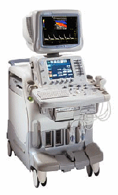
'The System of Choice for Shared Service. The LOGIQ® 7 system provides a full range of clinical applications including abdominal, small parts, surgery, vascular and cardiac imaging and the power of GE's patented TruScan architecture. Just imagine an ultrasound system so versatile and reliable that it can meet the demands of virtually any clinical setting. And an ergonomic design that improves scanning comfort and clinical work flow.'
Device Information and Specification
APPLICATIONS
Abdominal, cardiac, breast, intraoperative, musculoskeletal, neonatal, OB/GYN, orthopedic, pediatric, small parts, transcranial, urologic, vascular
CONFIGURATION
17' high resolution non-interlaced flat CRT, 4 active probe ports
B-mode, M-mode, coded harmonic imaging, color flow mode (CFM), power Doppler imaging (PDI), color Doppler, pulsed wave Doppler, tissue harmonic imaging
IMAGING OPTIONS
CrossXBeam spatial compounding, coded ultrasound acquisition),speckle reduction imaging (SRI), TruScan technology store raw data, CINE review with 4 speed types
OPTIONAL PACKAGE
Transesophageal scanning, stress echo, tissue velocity imaging (TVI), tissue velocity Doppler (TVD), contrast harmonic imaging
STORAGE, CONNECTIVITY, OS
Patient and image archive, HDD, DICOM 3.0, CD/DVD, MOD, Windows-based
DATA PROCESSING
Digital beamformer with 1024 system processing channel technology
H*W*D m (inch.)
1.62 * 0.61 * 0.99 (64 * 24 * 39)
WEIGHT
246 kg (498 lbs.)
POWER CONSUMPTION
less than 1.5 KVA
•  From GE Healthcare.;
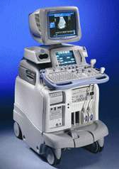
'The System of Choice for General Imaging Imagine a leading-edge ultrasound system so versatile that it can meet the demands of virtually any clinical setting. With the LOGIQ® 9, you'll have a high-performance system capable of multi-dimensional imaging for a full range of clinical applications - from abdominal to breast to vascular imaging. And an ergonomic design that improves scanning comfort and clinical work flow. Now, imagine what LOGIQ® 9 could do for you and your patients.'
Device Information and Specification
APPLICATIONS
Abdominal, cardiac, breast, intraoperative, musculoskeletal, neonatal, OB/GYN, orthopedic, pediatric, small parts, transcranial, urologic, vascular
CONFIGURATION
17' high resolution non-interlaced flat CRT, 4 active probe ports
B-mode, M-mode, coded harmonic imaging, color flow mode (CFM), power Doppler imaging (PDI), PW-HPRF, CW Doppler, color Doppler, pulsed wave Doppler, tissue harmonic imaging
IMAGING OPTIONS
CrossXBeam spatial compounding, coded ultrasound acquisition), speckle reduction imaging (SRI), TruScan technology store raw data, real-time 4D ultrasound, Tru 3D ultrasound
STORAGE, CONNECTIVITY, OS
Patient and image archive, HDD, DICOM 3.0, CD/DVD, MOD, PCMCIA, USB, Windows-based
DATA PROCESSING
Digital beamformer with 1024 system processing channel technology
H*W*D m (inch.)
1.62 * 0.61 * 0.99 (64 * 24 * 39)
WEIGHT
202 kg (408 lb.)
POWER CONSUMPTION
less than 2 KVA
•
(LUS) Diagnostic laparoscopy combined with laparoscopic ultrasound is used for staging tumors and to monitor surgical interventions like for example radiofrequency ablation or cryotherapy. Laparoscopic ultrasound provides direct contact imaging of organs with high frequency ultrasound. Laparoscopic ultrasound identifies and characterizes the tumor, guides the probe, and monitors the progression of the freezing or the thermal destruction. This procedure avoid unnecessary open surgery and improves selection of patients for tumor resection e.g., in liver and pancreas. Challenges of LUS are limitations of the intraoperative acoustic windows and the possible movement of the probe and that standard orientation techniques are difficult to apply with laparoscopic instruments, resulting in images from oblique planes. 3D ultrasound or special navigation systems may be helpful. See also Ultrasound Therapy. •  From ESAOTE S.p.A.;
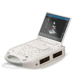
'The MyLab™30CV ultrasound system is an evolutionary step in ultrasound technology. Weighing less than 20 pounds, it is the first compact ultrasound system to deliver premium console performance. And with mobile, portable or stationary configurations, MyLab30CV can adapt to any clinical environment.'
Device Information and Specification
APPLICATIONS
Abdominal, breast, cardiac, OB/GYN, pediatric, pediatric cardiology, small parts, transcranial, vascular
CONFIGURATION
Portable
Linear: 4-10 MHz, convex: 2-5 MHz, phased: 1.6-10 MHz, micro convex: 5-7.5 MHz, endocavity: 5-7.5 MHz, pencil: 2 + 5 MHz
2-D, M-mode, duplex, triplex, color Doppler, pulsed wave Doppler, tissue velocity mapping (TVM), tissue enhancement imaging (TEI™), contrast harmonic imaging, stress echo, tissue velocity mapping for LV motion analysis (TVM), contrast tuned imaging for contrast media procedures (CnTI™), Qontrast™ for myocardium parameters quantification
STORAGE, CONNECTIVITY, OS
Digital patient archive/management, integrated CD/RW, RJ 45 and USB ports, Windows
H*W*D m (inch.)
0.16 * 0.36 * 0.50 (6.2 x 14 x 19.3)
WEIGHT
Less than 11 kg (20 lbs.)
•
Gynecologic ultrasound and obstetric ultrasound are two distinct applications of ultrasound imaging that serve different purposes in the field of women's health. While both involve the use of ultrasound technology to examine the pelvic region, they have different focuses and objectives.
Gynecologic [gynaecologic, Brit.] ultrasound primarily concentrates on the evaluation of the female reproductive organs, including the uterus, ovaries, fallopian tubes, and surrounding structures. It is commonly performed for various gynecological concerns, such as abnormal bleeding, pelvic pain, infertility investigations, and monitoring of reproductive disorders. It can identify signs of inflammation, the presence of free fluid, cysts, and tumors. This non-invasive technique aids in diagnosing and monitoring gynecological pathologies, facilitating early intervention and appropriate treatment. Typically, a transabdominal sonogram is performed with a full bladder to provide an initial assessment. However, if the pelvic ultrasound reveals any abnormalities or fails to provide a clear image of the organs, a more detailed evaluation can be achieved through a transvaginal sonography. This approach allows for improved visualization of the uterus and ovaries by placing the ultrasound probe inside the vagina. Obstetric ultrasound, also known as prenatal, fetal or pregnancy ultrasound, is the branch of medical imaging that focuses on the use of ultrasound technology to assess the health and development of a fetus during pregnancy. Women with uncomplicated pregnancies commonly undergo an ultrasound examination between the 16th and 20th week of gestation. This routine assessment, performed with a real-time scanner, serves to determine accurate gestational age, monitor fetal size, and assess overall growth. The middle of the pregnancy trimester provides a crucial window for detecting many abnormalities of fetal anatomy. Advanced imaging techniques enable healthcare professionals to identify potential structural issues. Early detection of these abnormalities allows for timely intervention, counseling, and the implementation of appropriate management strategies. See also Pregnancy Ultrasound, Pelvic Ultrasound, Hysterosalpingo Contrast Sonography and Vaginal Probe. Result Pages : |
Medical-Ultrasound-Imaging.com
former US-TIP.com
Member of SoftWays' Medical Imaging Group - MR-TIP • Radiology TIP • Medical-Ultrasound-Imaging
Copyright © 2008 - 2024 SoftWays. All rights reserved.
Terms of Use | Privacy Policy | Advertise With Us
former US-TIP.com
Member of SoftWays' Medical Imaging Group - MR-TIP • Radiology TIP • Medical-Ultrasound-Imaging
Copyright © 2008 - 2024 SoftWays. All rights reserved.
Terms of Use | Privacy Policy | Advertise With Us
[last update: 2023-11-06 01:42:00]


