Medical Ultrasound Imaging
Thursday, 9 May 2024
Info Sheets Out- side     | 'Fetal Ultrasound' p2 Searchterm 'Fetal Ultrasound' found in 19 articles 1 term [ • ] - 6 definitions [• ] - 12 booleans [• ]Result Pages : •
Diagnostic ultrasound imaging has no known risks or long-term side effects. Discomfort to the patient is very rare if the sonogram is accurately performed by using appropriate frequencies and intensity ranges. However, the application of the ALARA principle is always recommended.
There are reports of low birth weight of babies after applying more than the recommended ultrasound examinations during pregnancy. Women who think they might be pregnant should raise this issue with the doctor before undergoing an abdominal ultrasound, to avoid any harm to the fetus in the early stages of development. Since ultrasound is energy, sensitive tissues like the reproductive organs could possibly sustain damage if vibrated to a high degree by too intense ultrasound waves. In diagnostic ultrasonic procedures, such damage would only result from improper use of the equipment. Possible ultrasound bioeffects:
•
•
Due to increasing of temperature, dissolved gases from microbubbles come out of the contrast solution.
The thermal effect is controlled by the displayed thermal index and the mechanical index indicates the risk of cavitation. An ultrasound gel is applied to obtain better contact between the transducer and the skin. This has the consistency of thick mineral oil and is not associated with skin irritation or allergy. Specific conditions for which ultrasound may be selected as a treatment may be attached with higher risks. See also Ultrasound Imaging Procedures, Fetal Ultrasound and Obstetric and Gynecologic Ultrasound. • View NEWS results for 'Side Effect' (7). •
(TI) The definition of the thermal index is the ratio of the total acoustic power to that required raising a maximum temperature increase of 1 °C under defined assumptions. A thermal index of 1 indicates the acoustic power achieving a temperature increase of 1 °C. A thermal index of 2 has the doubled power but would not necessarily indicate a peak temperature rise of 2 °C. The temperature rise is dependent on tissue type and is particularly dependent on the presence of bone. Classifications of thermal indices:
•
TIS - thermal index soft tissue;
•
TIB - thermal index bone - bone at/near the focus;
•
TIC - thermal index cranial bone - bone at the surface.
For fetal ultrasound, the highest temperature increase would be expected occurring at bone. Therefore, TIB gives the worst-case conditions. If the ultrasound system can exceed an index of 1, the mechanical index and thermal index must be displayed. The displayed indices are based on the manufacturer's data. See also Cranial Bone Thermal Index, Bone Thermal Index, Soft Tissue Thermal Index. Further Reading: Basics:
News & More: •  From GE Healthcare.;
'GE is defining a new age of ultrasound. We call it Volume Ultrasound. GE's Voluson 730 Expert is a powerful system that enables real-time techniques for acquiring, navigating and analyzing volumetric images so that you can make clinical decisions with unprecedented confidence.'
Device Information and Specification
APPLICATIONS
Abdominal, breast, cardiac, musculoskeletal, neonatal, OB/GYN, pediatric, small parts, transcranial, urological, vascular
CONFIGURATION
15' high resolution non-interlaced flat CRT, 4 active probe ports
B-mode, M-mode, coded harmonic imaging (2-D), color flow mode (CFM), power Doppler imaging (PDI), color Doppler, pulsed wave Doppler, high pulse repetition frequency (HPRF) Doppler, tissue harmonic imaging, 3-D power Doppler
IMAGING OPTIONS
CrossXBeam spatial compounding, coded excitation , spatio-temporal image correlation (STIC), B-Flow (simultaneous imaging of tissue and blood flow), strain rate imaging (SRI)
OPTIONAL PACKAGE
STORAGE, CONNECTIVITY, OS
SonoView archiving and data management, network, HDD, DICOM 3.0, CD/DVD, MOD, USB, Windows-based
DATA PROCESSING
Digital beamformer with 512 system processing channel technology
H*W*D m (inch.)
1.43 * 0.69 * 1.02 (56 * 27 * 40)
WEIGHT
136 kg (300 lbs.)
• 
From SIUI Inc.;
Device Information and Specification
CONFIGURATION
Normal system, gray scale(256)
Linear and convex
PROBES STANDARD
2.5MHz ~ 11.0MHz, broad band, tri-frequency
IMAGING OPTIONS
Real zoom, max. Zoom * 4.0, multiplying power and position selectable
OPTIONAL PACKAGE
H*W*D m
1.30 * 0.52 * 0.77
WEIGHT
90kg (main unit)
POWER REQUIREMENT
AC 220V/110V, 50Hz/60Hz
POWER CONSUMPTION
0.45 KVA
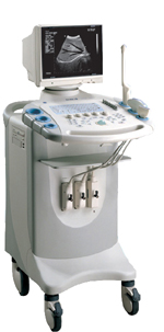
•  From SIUI Inc.;
'Dedicated to ultrasound industry, Shantou Institute of Ultrasonic Instruments, Inc. (SIUI) has launched Apogee 3500, the Digital Color Doppler Ultrasound Imaging System. With latest imaging technologies, high-definition image quality and excellent practical functions, the Apogee 3500 offers optimal solutions for clinical ultrasonic examination.' 'The Apogee 3500 is available with many high-density, super broadband and multi-frequency probes, such as convex, micro-convex, linear, vaginal, rectal and phased array probes, which are widely applied for different clinical diagnoses, including abdomen (liver, kidney, gall-bladder, pancreas), gynecology (uterus, ovary), obstetrics (early pregnancy, basic OB, complete OB, multi gestation, fetal echo), cardiology (adult and pediatric cardiology), small parts (thyroid, galactophore, testicles, neonate), peripheral vascular and prostate.'
Device Information and Specification
CONFIGURATION
Normal system, color - gray scale(256)
Linear, convex and phased array
PROBES STANDARD
2.0 MHz ~ 12.0 MHz, broad band, tri-frequency
B-mode (B, 2B, 4B), M-mode, B/M-mode, real-time compound imaging, panoramic imaging, trapezoidal imaging (linear probes), spectrum Doppler (PWD and CWD), color Doppler flow imaging (CDFI), color power angio (CPA), tissue harmonic imaging (THI)
IMAGING OPTIONS
Real-time ZOOM, zoom rate and position selectable
OPTIONAL PACKAGE
H*W*D m
1.29 * 0.52 * 0.75
WEIGHT
110 kg
POWER REQUIREMENT
AC 220V/110V, 50Hz/60Hz
POWER CONSUMPTION
0.6 KVA
Result Pages : | Share This Page Look Ups |
Medical-Ultrasound-Imaging.com
former US-TIP.com
Member of SoftWays' Medical Imaging Group - MR-TIP • Radiology TIP • Medical-Ultrasound-Imaging
Copyright © 2008 - 2024 SoftWays. All rights reserved.
Terms of Use | Privacy Policy | Advertise With Us
former US-TIP.com
Member of SoftWays' Medical Imaging Group - MR-TIP • Radiology TIP • Medical-Ultrasound-Imaging
Copyright © 2008 - 2024 SoftWays. All rights reserved.
Terms of Use | Privacy Policy | Advertise With Us
[last update: 2023-11-06 01:42:00]