Medical Ultrasound Imaging
Sunday, 20 September 2026

 Info Sheets  Out- side     | 'SD' p14 Searchterm 'SD' found in 199 articles 26 terms [ • ] - 173 definitions [• ] Result Pages : •
Echocardiography is the ultrasound examination of the heart. Depending on the used ultrasound system, echocardiograms can be two-dimensional slices or 3D real-time images of the heart. Based on the ultrasound principles the direction and speed of blood flow can be utilized e.g., to diagnose a leaking or stenosed valve or to identify intracardiac shunts. Different types of echocardiography:
•
•
contrast echocardiogram (CE);
The transthoracic echocardiogram (images are taken through the chest wall) is a non-invasive, highly accurate and quick assessment of the overall health of the heart. A more invasive method is to insert a specialized scope containing an echocardiography transducer (TEE probe) into the esophagus, and record images from there. The advantages are clearer images, since the transducer is closer to the heart. Contrast echocardiogram (CE) is already a valuable tool to delineate endocardial borders, direct invasive procedures, detect intracardiac shunts, assess myocardial perfusion and viability, and quantify coronary flow reserve and blood volumes (see also hemoglobin). The mechanism of microbubble CE is based on the physical principles of rarefaction and compression, leading to volume pulsations of microbubbles, and it is this change that results in CE signal. Stress echocardiograms are echocardiography exams used for detection of coronary artery disease. See also Diastole, Bicycle Stress Echocardiography, Resistive Index, and M-Mode Echocardiography.  Further Reading: Basics:
•
Sonography of the gallbladder is a reliable technique for diagnosing e.g., gallstones, cholecystitis, tumors, polyps, or ductal obstruction.
Patient should be examined with empty stomach and on a low fat diet the night before. Barium studies, endoscopy, ERCP, colonoscopy, and abdominal CT should be performed after this examination. Gallbladder ultrasound is best performed with a 5 MHz curved array or a linear array transducer in cases of a very superficial gallbladder. In obese patients or in patients with difficult sonographic access, a 3.5 MHz sector or curved linear transducer is advantageous. Gallbladder and biliary tree are usually imaged in supine and posterior oblique (LPO) positions. Sometimes very small gallstones are better visible in upright and prone position. Further Reading: Basics:
News & More:
• 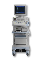 From Hitachi Medical Corporation (HMC), sales, marketing and service in the US by Hitachi Medical Systems America Inc.
The HI VISION™ 5500 - EUB-5500 fully digital ultrasound system delivers the latest generation of signal processing technology, sophisticated transducer design, and a host of features and options for advanced imaging capabilities across a wide range of clinical situations. This system is compatible with all Pentax ultrasound endoscopes.
Device Information and Specification
APPLICATIONS
Abdominal, brachytherapy/cryotherapy, breast, cardiac, dedicated biopsy, endoscopic, intraoperative, laparoscopic, musculoskeletal, OB/GYN, pediatric, small parts, urologic, vascular
CONFIGURATION
Compact system
Five frequency (except mini-probes)
Linear, convex, radial, miniradial/miniprobe, biplane, phased array, echoendoscope longitudinal, echoendoscope radial
3 modes of dynamic tissue harmonic imaging (dTHI), pulsed wave Doppler, continuous wave Doppler, color flow imaging, power Doppler, directional power Doppler, color flow angiography, real-time Doppler measurements
IMAGING OPTIONS
3RD generation color artifact suppression
OPTIONAL PACKAGE
STORAGE, CONNECTIVITY, OS
Patient and image database management system, HDD, FDD, MOD, CD-ROM, Network, DICOM 3.0, Windows XP
DATA PROCESSING
H*W*D m (inch.)
1.40 x 0.51 x 0.79 (55 x 20 x 31)
WEIGHT
130 kg (286 lbs.)
POWER CONSUMPTION
1.2kVA
ENVIRONMENTAL IMPACT
4096 btu/hr heat output
•
Usually, multiple probes are used because most transducers are only able to emit one frequency because the piezoelectric ceramic or crystals within it have a certain inherent frequency. Multi-frequency probes have multiple crystals with different frequencies and the desired specific frequency can be selected. Advanced probes can emit sound waves at different frequencies for the near and far fields. The disadvantage is that multi-frequency (multifrequency) probes have slower frame rates and therefore they are only useful for imaging of static structures. See also Dual Frequency Phased Array Transducer and Tri-Frequency Probe. •
The near field (also called Fresnel zone) is the proximal part of an ultrasound beam. The Fresnel zone is adjacent to the transducer surface and has a converging sound beam profile. A narrow beam shape is maintained in the near field owing to constructive and destructive interference patterns of sound wavelets emitted from the transducer crystal. The length of the near field is equal to r2/l = d2/4l where r is the radius, l is the ultrasound wavelength in the medium of propagation and d the diameter of the piezoelectric crystal. See also Beam Pattern, and Sonographic Features. Result Pages : | Share This Page     Look Ups |
 (.pdf)
Medical-Ultrasound-Imaging.com
former US-TIP.com
Member of SoftWays' Medical Imaging Group - MR-TIP • Radiology TIP • Medical-Ultrasound-Imaging
Copyright © 2008 - 2025 SoftWays. All rights reserved.
Terms of Use | Privacy Policy | Advertise With Us
former US-TIP.com
Member of SoftWays' Medical Imaging Group - MR-TIP • Radiology TIP • Medical-Ultrasound-Imaging
Copyright © 2008 - 2025 SoftWays. All rights reserved.
Terms of Use | Privacy Policy | Advertise With Us
[last update: 2025-04-05 02:38:00]