Medical Ultrasound Imaging
Sunday, 19 May 2024
Info Sheets Out- side     | 'Brachytherapy' p2 Searchterm 'Brachytherapy' found in 11 articles 1 term [ • ] - 10 definitions [• ] Result Pages : •  From Hitachi Medical Corporation (HMC), sales, marketing and service in the US by Hitachi Medical Systems America Inc.;
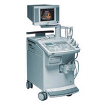
Powerful, flexible, and fast, the HI VISION™ 8500 - EUB-8500 diagnostic ultrasound scanner combines leading edge technologies with user-oriented operation for exceptional imaging and functionality. Available exclusively on the 8500, SonoElastography provides a new perspective on the physical properties of tumors and masses by determining and displaying the relative stiffness of tissue. Device Information and Specification
APPLICATIONS
Abdominal, brachytherapy/cryotherapy, breast, cardiac, dedicated biopsy, endoscopic, intraoperative, laparoscopic, musculoskeletal, OB/GYN, pediatric, small parts, urologic, vascular
CONFIGURATION
Compact system
RANGE OF PROBE TYPE
Linear, convex, radial, biplane, phased array, echoendoscope longitudinal, echoendoscope radial
PROBE FREQUENCIES
Linear: 5.0-13 MHz, convex: 2.5-7.5 MHz, phased:
2.0-7.5 MHz, sector: 2.0-7.5 MHz
4 Modes of dynamic tissue harmonic imaging (dTHI), pulsed wave Doppler, continuous wave Doppler, color flow imaging, power Doppler, directional power Doppler, color flow angiography, real-time Doppler measurements, quantitative tissue Doppler
IMAGING OPTIONS
HI COMPOUND imaging,
HI RES adaptive imaging, wideband pulse inversion imaging (WPI), Raw Data Freeze
OPTIONAL PACKAGE
IMAGING ENHANCEMENTS
3RD generation color artifact suppression
STORAGE, CONNECTIVITY, OS
Patient and image database management system, HDD, FDD, MOD, CD-ROM, Network, DICOM 3.0, Windows XP
DATA PROCESSING
Octal beam processing, 12 bit Gigasampling A/D for precise signal reproduction
H*W*D m (inch.)
1.50 * 0.56 * 1.02 (59 x 22 x 40)
WEIGHT
159 kg (351 lbs.)
POWER CONSUMPTION
1.5kVA
•
From Hitachi Medical Corporation (HMC);The HI VISION™ 6500 - EUB-6500 high resolution digital ultrasound system offers advanced clinical imaging, enhanced operating efficiency, and remarkable clinical flexibility, all in robust and versatile configuration that simply represents a better clinical solution in a variety of real-world, real-work arenas.
Device Information and Specification
APPLICATIONS
Abdominal, brachytherapy/cryotherapy, breast, cardiac, dedicated biopsy, endoscopic, intraoperative, laparoscopic, musculoskeletal, OB/GYN, pediatric, small parts, urologic, vascular
CONFIGURATION
Compact system
Linear, convex, radial, miniradial/miniprobe, biplane, phased array, echoendoscope longitudinal, echoendoscope radial
Tissue Doppler imaging (TDI), pulsed wave Doppler, continuous wave Doppler, color flow imaging, power Doppler, directional power Doppler, color flow angiography, real-time Doppler measurements, 4 modes of dynamic tissue harmonic imaging (dTHI), wideband pulse inversion imaging (WPI)
IMAGING OPTIONS
3RD generation color artifact suppression
OPTIONAL PACKAGE
3D ultrasound, dual omni-directional M-mode display, steerable CW Doppler, dynamic contrast harmonics imaging, stress echo, Pentax EUS and Fujinon Mini-probe
STORAGE, CONNECTIVITY, OS
Patient and image database management system, HDD, FDD, MOD, CD-ROM, Network, DICOM 3.0, Windows XP
DATA PROCESSING
H*W*D m (inch.)
1.40 x 0.51 x 0.79 (55 x 20 x 31)
WEIGHT
130 kg (286 lbs.)
POWER CONSUMPTION
1.2kVA
ENVIRONMENTAL POLLUTION
4096 btu/hr heat output
•
The prostate is a walnut-shaped gland surrounding the beginning of the urethra in front of the rectum and below the bladder. The prostate can become enlarged (particularly in men over age 50) and develop diseases like prostate cancer or inflammation (prostatitis). A large tumor can be felt by a rectal examination. The most effective way of detecting the early signs of prostate cancer is a combination of a prostate-specific antigen (PSA) blood test and a prostate ultrasound examination. An abnormally high level of PSA can indicate prostate cancer or other prostate diseases such as benign prostatic hypertrophy or prostatitis. The transrectal sonography is an important diagnostic ultrasound procedure in determining whether there is any benign enlargement of the prostate or any abnormal nodules. The imaging is performed with a rectal probe, yielding high resolution. High resolution 3D ultrasound provides reliable and accurate determination of the size and the location of cancer. Additionally, ultrasound elastography is a technique in development to improve the specificity and sensitivity of cancer detection. Ultrasound is also used to detect whether cancerous tissue is still only within the prostate or whether it has begun to spread out and to guide a diagnostic biopsy or ultrasound therapy. See also Brachytherapy, and High Intensity Focused Ultrasound. Further Reading: News & More:
•  From Siemens Medical Systems;
'This extremely flexible system supports targeted applications for OB/GYN, Radiology, Internal Medicine, Urology, and Prostate Brachytherapy. The large transducer selection, most with high-frequency capabilities, provides the right tool for general imaging, radiology, and internal medicine applications.'
Device Information and Specification
CLINICAL APPLICATION
OB/GYN, cerebrovascular, orthopedics, urology, peripheral vascular, pediatrics, small parts, surgery, abdominal, musculoskeletal
CONFIGURATION
Mobile, compact
DISPLAY MODES
Broadband, high-fidelity, multi-frequency, linear and curved
PROBE PORTS
Two - four
256
MEASUREMENT/CALCULATION FUNCTIONS
OB/GYN measurements and report package, off-line analysis
OPTIONAL PACKAGE
IMAGE PROCESSING
•
(TRUS) Transrectal sonography (also called transrectal ultrasonography, transrectal echography (TRE), endorectal ultrasound (ERUS or EUS)) is an ultrasound procedure used to examine the prostate gland, the rectum or bladder. A small, lubricated transducer placed into the rectum releases sound waves, which create echoes as they enter the region of interest. A computer creates a picture called a sonogram. TRUS is commonly used for guidance during a prostate needle biopsy and may be used to deliver brachytherapy and monitor cancer treatment. Transrectal ultrasonography detects enlargement, tumors and other abnormalities of the prostate, rectal polyps, rectal cancer, perianal infection, and sphincter muscle injuries. TRUS is also performed on male patients with infertility to view the prostate and surrounding structures and on patients with suspected bladder conditions or disease to view the bladder. See also Transurethral Sonography, Endoscopic Ultrasound, Pelvic Ultrasound, Rectal Probe, Biplane Probe, Endocavitary Echography and High Intensity Focused Ultrasound. Further Reading: News & More:
Result Pages : | Share This Page Look Ups |
Medical-Ultrasound-Imaging.com
former US-TIP.com
Member of SoftWays' Medical Imaging Group - MR-TIP • Radiology TIP • Medical-Ultrasound-Imaging
Copyright © 2008 - 2024 SoftWays. All rights reserved.
Terms of Use | Privacy Policy | Advertise With Us
former US-TIP.com
Member of SoftWays' Medical Imaging Group - MR-TIP • Radiology TIP • Medical-Ultrasound-Imaging
Copyright © 2008 - 2024 SoftWays. All rights reserved.
Terms of Use | Privacy Policy | Advertise With Us
[last update: 2023-11-06 01:42:00]