Medical Ultrasound Imaging
Sunday, 19 May 2024
Info Sheets Out- side     | 'Continuous Wave Doppler' p3 Searchterm 'Continuous Wave Doppler' found in 18 articles 1 term [ • ] - 14 definitions [• ] - 3 booleans [• ]Result Pages : •
Spectral Doppler refers to the combination of either continuous wave Doppler or pulsed Doppler with a spectral display. Spectral Doppler provides a quantitative analysis of the velocity and direction of blood flow. The Fourier spectrum analyzer performs a fast Fourier transformation on the Doppler signal. The amplitudes of the resulting spectra are encoded as brightness. In the 2D spectral display, the frequency shift is depicted in the vertical and the time in the horizontal axis. The range of blood velocities in the volume produces a corresponding range of frequency shifts. See also Acceleration Index and Triplex Exam. •
Ultrasound biomicroscopy utilizes high frequency (10 - 50 MHz) diagnostic ultrasound to examine living tissue at a microscopic level and allows to image the skin with extremely high resolution to a depth of 2-3 centimeters. Ultrasound biomicroscopy images provide detailed anatomical information that can lead to better and more accurate treatments and avoid a biopsy. Ultrasound biomicroscopy improves also the spatial resolution of US images of the anterior segment of the eye. US biomicroscopy of the eye operates in the 50 MHz range with a possible axial resolution on the order of 30 μm. In this frequency range, tissue penetration of only approximately 5 mm is attainable. Both continuous wave Doppler and high-frequency pulsed Doppler can be used. See also Ultrasound Imaging Procedures, A-Scan, B-Scan and C-Scan. Further Reading: News & More:
•
Ultrasound imaging is excellent for diagnosing cysts and other fluids in soft tissue. For ultrasound imaging or ultrasonography, different modes are used to examine the arterial/venous system, heart, pancreas, urinary system, ovaries, spinal cord, joints and more. Power levels, frequencies used, amplification, and beamforming determine the clarity of the image. These things are controlled by the sonographer, interacting with the properties of the ultrasound machine. Various imaging modes:
•
•
•
•
•
•  From GE Healthcare.;
'Vivid 7 Dimension, a premier cardiovascular ultrasound system from GE Healthcare, expands on the strength of a powerful imaging platform to offer new, innovative technology of dimensional proportions.'
Device Information and Specification
CONFIGURATION
Multi-frequency, linear, convex, phased, sector
B-mode, C-mode, M-mode (and 2-D), triplex mode, harmonic imaging, color flow mapping, 3D ultrasound display, power Doppler imaging (PDI), color Doppler, pulsed wave Doppler, continuous wave Doppler, tissue velocity imaging (TVI), tissue type imaging (TTI), strain rate imaging (SRI), tissue synchronization imaging (TSI)
IMAGING OPTIONS
CINE review with 5 speed types, bi- andtri-plane imaging with e.g. stress echo and tissue synchronization imaging
STORAGE, CONNECTIVITY, OS
Patient and image archive, HDD, MOD, DVD, USB flash card, DICOM 3.0 Windows-based
DATA PROCESSING
Digital beamformer with 1024 system processing channel technology
H*W*D m (inch.)
1.58 * 0.64 * 0.89 (62 * 25 * 35)
WEIGHT
191 kg (420 lbs.)
POWER CONSUMPTION
less than 2 KVA
• 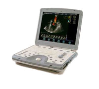 From GE Healthcare.;
'The incredible Vivid i system establishes a completely new level of cardiovascular performance that gives clinicians the freedom to get diagnostic results outside of the echo lab.'
Device Information and Specification
APPLICATIONS
CONFIGURATION
Notebook
M-mode (and 2-D), triplex mode, harmonic imaging, color flow mapping, pulsed wave Doppler, continuous wave Doppler, power Doppler, color Doppler, tissue harmonic imaging, color flow mapping
IMAGING OPTIONS
STORAGE, CONNECTIVITY, OS
Patient and image archive, HDD, DICOM, CD/DVD, MOD, USB flash, PCMCIA, eVue for remote monitoring, MPEGvue foruniversal record sharing
H*W*D cm (inch.)
7 * 36 * 32 (2.6 x 14.1 x 12.3)
WEIGHT
5 kg (11 lbs.)
POWER CONSUMPTION
Rechargeable battery provides up to 1.0 hour of full scan operation
Result Pages : | Share This Page Look Ups |
Medical-Ultrasound-Imaging.com
former US-TIP.com
Member of SoftWays' Medical Imaging Group - MR-TIP • Radiology TIP • Medical-Ultrasound-Imaging
Copyright © 2008 - 2024 SoftWays. All rights reserved.
Terms of Use | Privacy Policy | Advertise With Us
former US-TIP.com
Member of SoftWays' Medical Imaging Group - MR-TIP • Radiology TIP • Medical-Ultrasound-Imaging
Copyright © 2008 - 2024 SoftWays. All rights reserved.
Terms of Use | Privacy Policy | Advertise With Us
[last update: 2023-11-06 01:42:00]