Medical Ultrasound Imaging
Monday, 20 May 2024
Info Sheets Out- side     | 'Duplex' p2 Searchterm 'Duplex' found in 10 articles 2 terms [ • ] - 8 definitions [• ] Result Pages : •
Compound B-mode imaging takes different forms and refers to different methods of creating the ultrasound image. Real-time compound ultrasound improves the image quality of B-mode scanning by combining ultrasound information obtained from multiple angles. The used averaging process of compound B-mode reduces artifacts and improves the representation of true image data. B-mode images and Doppler mode images (see also Duplex) can be compounded on the display to improve the visualization of the anatomical relationships between vessels and the surrounding tissues. Further Reading: News & More:
•  From ESAOTE S.p.A.;
'The MyLab™30CV ultrasound system is an evolutionary step in ultrasound technology. Weighing less than 20 pounds, it is the first compact ultrasound system to deliver premium console performance. And with mobile, portable or stationary configurations, MyLab30CV can adapt to any clinical environment.'
Device Information and Specification
APPLICATIONS
Abdominal, breast, cardiac, OB/GYN, pediatric, pediatric cardiology, small parts, transcranial, vascular
CONFIGURATION
Portable
Linear: 4-10 MHz, convex: 2-5 MHz, phased: 1.6-10 MHz, micro convex: 5-7.5 MHz, endocavity: 5-7.5 MHz, pencil: 2 + 5 MHz
2-D, M-mode, duplex, triplex, color Doppler, pulsed wave Doppler, tissue velocity mapping (TVM), tissue enhancement imaging (TEI™), contrast harmonic imaging, stress echo, tissue velocity mapping for LV motion analysis (TVM), contrast tuned imaging for contrast media procedures (CnTI™), Qontrast™ for myocardium parameters quantification
STORAGE, CONNECTIVITY, OS
Digital patient archive/management, integrated CD/RW, RJ 45 and USB ports, Windows
H*W*D m (inch.)
0.16 * 0.36 * 0.50 (6.2 x 14 x 19.3)
WEIGHT
Less than 11 kg (20 lbs.)
•
Ultrasound imaging is excellent for diagnosing cysts and other fluids in soft tissue. For ultrasound imaging or ultrasonography, different modes are used to examine the arterial/venous system, heart, pancreas, urinary system, ovaries, spinal cord, joints and more. Power levels, frequencies used, amplification, and beamforming determine the clarity of the image. These things are controlled by the sonographer, interacting with the properties of the ultrasound machine. Various imaging modes:
•
•
•
•
•
•
Vascular ultrasound obtains images and measures blood flow velocity in the carotids, abdominal aorta, and vessels of kidneys, arms, or legs. Blockages in arteries, blood clots in veins, or abdominal aortic aneurysm can be detected. These abnormalities in blood flow are usually examined with different Doppler techniques. In addition, the speed and direction of blood flow can be color coded in a color map. Duplex techniques show both, the vessels and the surrounding tissue. The use of ultrasound contrast agents improves the left ventricular opacification in cardiac ultrasound examination. Usually, for a vascular ultrasound no special preparation is needed. See also Echocardiography, Venous Ultrasound, Adventitia, Intima, Temporal Mean Velocity, and Intravascular Ultrasound. Further Reading: News & More:
• 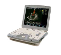 From GE Healthcare.;
'The incredible Vivid i system establishes a completely new level of cardiovascular performance that gives clinicians the freedom to get diagnostic results outside of the echo lab.'
Device Information and Specification
APPLICATIONS
CONFIGURATION
Notebook
M-mode (and 2-D), triplex mode, harmonic imaging, color flow mapping, pulsed wave Doppler, continuous wave Doppler, power Doppler, color Doppler, tissue harmonic imaging, color flow mapping
IMAGING OPTIONS
STORAGE, CONNECTIVITY, OS
Patient and image archive, HDD, DICOM, CD/DVD, MOD, USB flash, PCMCIA, eVue for remote monitoring, MPEGvue foruniversal record sharing
H*W*D cm (inch.)
7 * 36 * 32 (2.6 x 14.1 x 12.3)
WEIGHT
5 kg (11 lbs.)
POWER CONSUMPTION
Rechargeable battery provides up to 1.0 hour of full scan operation
Result Pages : | Share This Page Look Ups |
Medical-Ultrasound-Imaging.com
former US-TIP.com
Member of SoftWays' Medical Imaging Group - MR-TIP • Radiology TIP • Medical-Ultrasound-Imaging
Copyright © 2008 - 2024 SoftWays. All rights reserved.
Terms of Use | Privacy Policy | Advertise With Us
former US-TIP.com
Member of SoftWays' Medical Imaging Group - MR-TIP • Radiology TIP • Medical-Ultrasound-Imaging
Copyright © 2008 - 2024 SoftWays. All rights reserved.
Terms of Use | Privacy Policy | Advertise With Us
[last update: 2023-11-06 01:42:00]