Medical Ultrasound Imaging
Sunday, 19 May 2024
Info Sheets Out- side     | 'Longitudinal Wave' Searchterm 'Longitudinal Wave' found in 6 articles 1 term [ • ] - 2 definitions [• ] - 3 booleans [• ]Result Pages : • Longitudinal Wave
A longitudinal wave is a waveform in which the particle motion is parallel to the direction of the wave travel. Sound propagates as longitudinal waves. A series of longitudinal waves make up the ultrasound beam.
A longitudinal wave is produced when a piezoelectric ceramic in an ultrasound transducer, transmits it's back and forth oscillation into a continuous, elastic medium. The particles of the medium are made to oscillate in the direction of the wave propagation, but are otherwise stationary. •
(TIB) The bone thermal index is an exposure model for the case that the ultrasound beam passes through soft tissue and a focal region is in the immediate vicinity of bone. The longitudinal waves of ultrasound are reflected and transformed into transverse waves, creating a heating effect. Muscle and bone absorb more energy at interfaces with other heterogeneous tissues. See also Sheer Wave. •
(short for ultrasound beam) The sound beam is the confined, directional beam of ultrasound traveling as a longitudinal wave from the transducer face into the propagation medium. The near field and the far field are two separate regions along the beam. Sound beams are either steered mechanically or electrically. Both rapidly sweep sound waves through tissues. See also Sheer Wave, Beam Vessel Angle, Beam Steering, and Huygens Principle. • 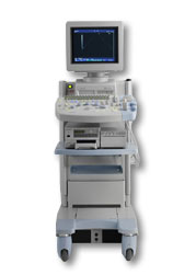 From Hitachi Medical Corporation (HMC), sales, marketing and service in the US by Hitachi Medical Systems America Inc.
The HI VISION™ 5500 - EUB-5500 fully digital ultrasound system delivers the latest generation of signal processing technology, sophisticated transducer design, and a host of features and options for advanced imaging capabilities across a wide range of clinical situations. This system is compatible with all Pentax ultrasound endoscopes.
Device Information and Specification
APPLICATIONS
Abdominal, brachytherapy/cryotherapy, breast, cardiac, dedicated biopsy, endoscopic, intraoperative, laparoscopic, musculoskeletal, OB/GYN, pediatric, small parts, urologic, vascular
CONFIGURATION
Compact system
Five frequency (except mini-probes)
Linear, convex, radial, miniradial/miniprobe, biplane, phased array, echoendoscope longitudinal, echoendoscope radial
3 modes of dynamic tissue harmonic imaging (dTHI), pulsed wave Doppler, continuous wave Doppler, color flow imaging, power Doppler, directional power Doppler, color flow angiography, real-time Doppler measurements
IMAGING OPTIONS
3RD generation color artifact suppression
OPTIONAL PACKAGE
STORAGE, CONNECTIVITY, OS
Patient and image database management system, HDD, FDD, MOD, CD-ROM, Network, DICOM 3.0, Windows XP
DATA PROCESSING
H*W*D m (inch.)
1.40 x 0.51 x 0.79 (55 x 20 x 31)
WEIGHT
130 kg (286 lbs.)
POWER CONSUMPTION
1.2kVA
ENVIRONMENTAL IMPACT
4096 btu/hr heat output
•  From Hitachi Medical Corporation (HMC), sales, marketing and service in the US by Hitachi Medical Systems America Inc.;
Powerful, flexible, and fast, the HI VISION™ 8500 - EUB-8500 diagnostic ultrasound scanner combines leading edge technologies with user-oriented operation for exceptional imaging and functionality. Available exclusively on the 8500, SonoElastography provides a new perspective on the physical properties of tumors and masses by determining and displaying the relative stiffness of tissue. Device Information and Specification
APPLICATIONS
Abdominal, brachytherapy/cryotherapy, breast, cardiac, dedicated biopsy, endoscopic, intraoperative, laparoscopic, musculoskeletal, OB/GYN, pediatric, small parts, urologic, vascular
CONFIGURATION
Compact system
RANGE OF PROBE TYPE
Linear, convex, radial, biplane, phased array, echoendoscope longitudinal, echoendoscope radial
PROBE FREQUENCIES
Linear: 5.0-13 MHz, convex: 2.5-7.5 MHz, phased:
2.0-7.5 MHz, sector: 2.0-7.5 MHz
4 Modes of dynamic tissue harmonic imaging (dTHI), pulsed wave Doppler, continuous wave Doppler, color flow imaging, power Doppler, directional power Doppler, color flow angiography, real-time Doppler measurements, quantitative tissue Doppler
IMAGING OPTIONS
HI COMPOUND imaging,
HI RES adaptive imaging, wideband pulse inversion imaging (WPI), Raw Data Freeze
OPTIONAL PACKAGE
IMAGING ENHANCEMENTS
3RD generation color artifact suppression
STORAGE, CONNECTIVITY, OS
Patient and image database management system, HDD, FDD, MOD, CD-ROM, Network, DICOM 3.0, Windows XP
DATA PROCESSING
Octal beam processing, 12 bit Gigasampling A/D for precise signal reproduction
H*W*D m (inch.)
1.50 * 0.56 * 1.02 (59 x 22 x 40)
WEIGHT
159 kg (351 lbs.)
POWER CONSUMPTION
1.5kVA
Result Pages : | Share This Page Look Ups |
Medical-Ultrasound-Imaging.com
former US-TIP.com
Member of SoftWays' Medical Imaging Group - MR-TIP • Radiology TIP • Medical-Ultrasound-Imaging
Copyright © 2008 - 2024 SoftWays. All rights reserved.
Terms of Use | Privacy Policy | Advertise With Us
former US-TIP.com
Member of SoftWays' Medical Imaging Group - MR-TIP • Radiology TIP • Medical-Ultrasound-Imaging
Copyright © 2008 - 2024 SoftWays. All rights reserved.
Terms of Use | Privacy Policy | Advertise With Us
[last update: 2023-11-06 01:42:00]