Medical Ultrasound Imaging
Saturday, 11 May 2024
Info Sheets Out- side     | 'Noise' p2 Searchterm 'Noise' found in 15 articles 2 terms [ • ] - 13 definitions [• ] Result Pages : •
(THI) Tissue harmonic imaging (also called native harmonic imaging) is a signal processing technique which addresses ultrasound limitations like penetration and resolution. Tissue harmonic imaging reduces noise and clutter by improving signal to noise ratio and resolution. The signal penetration in soft tissue increases as the transmit frequency is decreased, by simultaneous decreased image resolution. As an ultrasound wave propagates through the target media a change occurs in the shape and frequency of the transmitted signal. The change is due to the normal resistance of tissue to propagate sound energy. This resistance and the resulting signal change is called a harmonic oscillation. For harmonic imaging the input frequency doubles the output frequency, for example a transmit frequency of 3.0 MHz. which would provide maximum penetration will return a harmonic frequency of 6.0 MHz. The returning higher frequency signal has to only travel one direction to the probe. The advantages of high frequency imaging and the one-way travel effect are decreased reverberation, beam aberration, and side lobes, as well as increased resolution and cystic clearing. •
(ADC) A system that receives analog input data and produces digital values at its output. Analog to digital converters are used by the ultrasound scanner to convert the received signal into a format more compatible with the computer systems. Ultrasound front-end-systems as well as many others sophisticated electronic systems use these analog signal processing components in connection with e.g., low-noise amplifier (LNA), and time gain compensation amplifier (TGC) as key elements in determining the overall system performance. See also Digital to Analog Converter, and Digitization. •
The wider the ultrasound beam, the more severe the problem with volume averaging and the beam-width artifact, to avoid this, the ultrasound beam can be shaped with lenses.
Different possibilities to focus the beam:
•
Mechanical focusing is performed by placing an acoustic lens on the surface of the transducer or using a transducer with a concave face.
•
Electronic focusing uses multiple phased array (annular or linear) elements, sequentially fired to focus the beam.
Conventional multi-element transducers are electronically focused in order to minimize beam width. This transducer type can be focused electronically only along the long axis of the probe where there are multiple elements, along the short axis (elevation axis) are conventional transducers only one element wide. Electronic focusing in any axis requires multiple transducer elements arrayed along that axis. Short axis focusing of conventional multi-element transducers requires an acoustic lens which has a fixed focal length. For operation at frequencies at or even above 10 MHz, quantization noise reduces contrast resolution. Digital beamforming gives better control over time delay quantization errors. In digital beamformers the delay accuracy is improved, thus allowing higher frequency operation. In analog beamformers, delay accuracy is in the order of 20 ns. Phased beamformers are suitable to handle linear phased arrays and are used for sector formats such as required in cardiography to improve image quality. Beamforming in ultrasound instruments for medical imaging uses analog delay lines. The signal from each individual element is delayed in order to steer the beam in the desired direction and focuses the beam. The receive beamformer tracks the depth and focuses the receive beam as the depth increases for each transmitted pulse. The receive aperture increase with depth. The lateral resolution is constant with depth, and decreases the sensitivity to aberrations in the imaged tissue. A requirement for dynamic control of the used elements is given. Since often a weighting function (apodization) is used for side lobe reduction, the element weights also have to be dynamically updated with depth. See also Huygens Principle. • 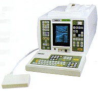 From SIUI Inc.;
'The SIUI CTS-200 is one of few 'linear only' units to be developed with a digital scan converter to process all incoming signals. With a digital processor, not only is the incoming signal processed faster but less noise is introduced into the signal path. The images are cleaner, crisper than you have experienced, even in machines at many times the price.' 'CTS-200 is suitable to a wide range of examination of liver, gallbladder, kidney, spleen, pancreas, thyroid gland, breast, uterus, urinary bladder, OB/GYN, etc. It is a portable ultrasound scanner of high performance.'
Device Information and Specification
APPLICATIONS
See description above
CONFIGURATION
Portable, gray scale(256)
Linear
PROBES STANDARD
1 * 3.5MHz Linear Probe EZU-PL21
PROBES FREQUENCY
3.5MHz, 7.5MHz
IMAGING OPTIONS
*1, *1.5, *2 as well as depth shift
OPTIONAL PACKAGE
7.5MHz Linear Probe EZU-PL23; 7.5MHz Transrectal Probe EUP-U23; 3.5MHz Biopsy Probe EUP-B11A; Trolley/Mobile Cart; ...
DATA PROCESSING
Pre-processing, correlation-processing, interpolation
POWER REQUIREMENT
AC 220V/110V, 50Hz/60Hz
POWER CONSUMPTION
0.05 KVA
•
(CEUS) Contrast agents increase the reflection of ultrasonic energy, improve the signal to noise ratio and caused by that the detection of abnormal microvascular and macrovascular disorders. Contrast enhanced ultrasound is used in abdominal ultrasound (liver sonography) as well as in cerebrovascular examinations e.g., for an accurate grading of carotid stenosis. The used contrast agents are safe and well tolerated. The quality of the enhancement depends on:
•
the concentration of the contrast agent;
•
the type of injection, flow rate;
•
the patient characteristics;
•
the microbubble quality and properties of the filling gas and the shell.
The additional use of ultrasound contrast agents (USCAs) may overcome typical limitations like poor contrast of B-mode imaging or limited sensitivity of Doppler techniques. The development of new ultrasound applications (e.g., blood flow imaging, perfusion quantification) depends also from the development of pulse sequences for bubble specific imaging. In addition, contrast enhanced ultrasound improves the monitoring of ultrasound guided interventions like RF thermal ablation. See also Contrast Enhanced Doppler Imaging, Contrast Harmonic Imaging, Contrast Imaging Techniques and Contrast Pulse Sequencing. Further Reading: News & More:
Result Pages : | Share This Page Look Ups |
Medical-Ultrasound-Imaging.com
former US-TIP.com
Member of SoftWays' Medical Imaging Group - MR-TIP • Radiology TIP • Medical-Ultrasound-Imaging
Copyright © 2008 - 2024 SoftWays. All rights reserved.
Terms of Use | Privacy Policy | Advertise With Us
former US-TIP.com
Member of SoftWays' Medical Imaging Group - MR-TIP • Radiology TIP • Medical-Ultrasound-Imaging
Copyright © 2008 - 2024 SoftWays. All rights reserved.
Terms of Use | Privacy Policy | Advertise With Us
[last update: 2023-11-06 01:42:00]