Medical Ultrasound Imaging
Monday, 20 May 2024
Info Sheets Out- side     | 'Window' p3 Searchterm 'Window' found in 26 articles 6 terms [ • ] - 20 definitions [• ] Result Pages : •
Handheld ultrasound systems are portable devices for smartphone or tablet and are increasingly common in emergency, intensive care and veterinary medicine, but also in the pocket of the stationary doctor. This type of ultrasound machine enables immediate diagnoses directly on site (handheld point-of-care-ultrasound / HPOCUS) and quickly provide information regarding the patient's further care. Handheld ultrasound machines fit into a single-use plastic cover and can be easily disinfected, making them particularly useful in infectious environments. The most striking advantage of handheld POCUS devices is the small footprint. The design is very compact, lightweight (approx. 200 g/0,44 lbs. - 500 g/1,1 lbs) and flexible. Due to this compactness and the necessary technical compression, the quality of the imaging is still limited compared to 'high-end devices', but sufficient to the extent that handheld ultrasound devices are already successfully used in many medical disciplines. Depending on the model, handheld ultrasound systems run on Android, iOS, Windows or proprietary operating systems. They are connected to the end device via USB cable or wirelessly via Bluetooth or WiFi. The respective end device is used as an ultrasound monitor to display the ultrasound images. The associated app is operated via touchscreen, although some devices have a few buttons, e.g. for recording ultrasound images or freeze images. The images can be stored and managed on the end device itself, the inserted memory card or in the cloud. Theoretically, also a private smartphone can be connected, but this can lead to complications with reimbursement. See also Portable Ultrasound Machine, Ultrasound Technology, Environmental Protection, Ultrasound Accessories and Supplies and Sonographer. Further Reading: News & More:
• 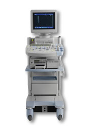 From Hitachi Medical Corporation (HMC), sales, marketing and service in the US by Hitachi Medical Systems America Inc.
The HI VISION™ 5500 - EUB-5500 fully digital ultrasound system delivers the latest generation of signal processing technology, sophisticated transducer design, and a host of features and options for advanced imaging capabilities across a wide range of clinical situations. This system is compatible with all Pentax ultrasound endoscopes.
Device Information and Specification
APPLICATIONS
Abdominal, brachytherapy/cryotherapy, breast, cardiac, dedicated biopsy, endoscopic, intraoperative, laparoscopic, musculoskeletal, OB/GYN, pediatric, small parts, urologic, vascular
CONFIGURATION
Compact system
Five frequency (except mini-probes)
Linear, convex, radial, miniradial/miniprobe, biplane, phased array, echoendoscope longitudinal, echoendoscope radial
3 modes of dynamic tissue harmonic imaging (dTHI), pulsed wave Doppler, continuous wave Doppler, color flow imaging, power Doppler, directional power Doppler, color flow angiography, real-time Doppler measurements
IMAGING OPTIONS
3RD generation color artifact suppression
OPTIONAL PACKAGE
STORAGE, CONNECTIVITY, OS
Patient and image database management system, HDD, FDD, MOD, CD-ROM, Network, DICOM 3.0, Windows XP
DATA PROCESSING
H*W*D m (inch.)
1.40 x 0.51 x 0.79 (55 x 20 x 31)
WEIGHT
130 kg (286 lbs.)
POWER CONSUMPTION
1.2kVA
ENVIRONMENTAL IMPACT
4096 btu/hr heat output
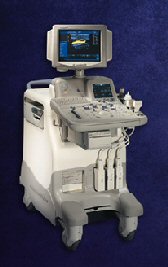
•  From Hitachi Medical Corporation (HMC), sales, marketing and service in the US by Hitachi Medical Systems America Inc.;
Powerful, flexible, and fast, the HI VISION™ 8500 - EUB-8500 diagnostic ultrasound scanner combines leading edge technologies with user-oriented operation for exceptional imaging and functionality. Available exclusively on the 8500, SonoElastography provides a new perspective on the physical properties of tumors and masses by determining and displaying the relative stiffness of tissue. Device Information and Specification
APPLICATIONS
Abdominal, brachytherapy/cryotherapy, breast, cardiac, dedicated biopsy, endoscopic, intraoperative, laparoscopic, musculoskeletal, OB/GYN, pediatric, small parts, urologic, vascular
CONFIGURATION
Compact system
RANGE OF PROBE TYPE
Linear, convex, radial, biplane, phased array, echoendoscope longitudinal, echoendoscope radial
PROBE FREQUENCIES
Linear: 5.0-13 MHz, convex: 2.5-7.5 MHz, phased:
2.0-7.5 MHz, sector: 2.0-7.5 MHz
4 Modes of dynamic tissue harmonic imaging (dTHI), pulsed wave Doppler, continuous wave Doppler, color flow imaging, power Doppler, directional power Doppler, color flow angiography, real-time Doppler measurements, quantitative tissue Doppler
IMAGING OPTIONS
HI COMPOUND imaging,
HI RES adaptive imaging, wideband pulse inversion imaging (WPI), Raw Data Freeze
OPTIONAL PACKAGE
IMAGING ENHANCEMENTS
3RD generation color artifact suppression
STORAGE, CONNECTIVITY, OS
Patient and image database management system, HDD, FDD, MOD, CD-ROM, Network, DICOM 3.0, Windows XP
DATA PROCESSING
Octal beam processing, 12 bit Gigasampling A/D for precise signal reproduction
H*W*D m (inch.)
1.50 * 0.56 * 1.02 (59 x 22 x 40)
WEIGHT
159 kg (351 lbs.)
POWER CONSUMPTION
1.5kVA
•
From Hitachi Medical Corporation (HMC);The HI VISION™ 6500 - EUB-6500 high resolution digital ultrasound system offers advanced clinical imaging, enhanced operating efficiency, and remarkable clinical flexibility, all in robust and versatile configuration that simply represents a better clinical solution in a variety of real-world, real-work arenas.
Device Information and Specification
APPLICATIONS
Abdominal, brachytherapy/cryotherapy, breast, cardiac, dedicated biopsy, endoscopic, intraoperative, laparoscopic, musculoskeletal, OB/GYN, pediatric, small parts, urologic, vascular
CONFIGURATION
Compact system
Linear, convex, radial, miniradial/miniprobe, biplane, phased array, echoendoscope longitudinal, echoendoscope radial
Tissue Doppler imaging (TDI), pulsed wave Doppler, continuous wave Doppler, color flow imaging, power Doppler, directional power Doppler, color flow angiography, real-time Doppler measurements, 4 modes of dynamic tissue harmonic imaging (dTHI), wideband pulse inversion imaging (WPI)
IMAGING OPTIONS
3RD generation color artifact suppression
OPTIONAL PACKAGE
3D ultrasound, dual omni-directional M-mode display, steerable CW Doppler, dynamic contrast harmonics imaging, stress echo, Pentax EUS and Fujinon Mini-probe
STORAGE, CONNECTIVITY, OS
Patient and image database management system, HDD, FDD, MOD, CD-ROM, Network, DICOM 3.0, Windows XP
DATA PROCESSING
H*W*D m (inch.)
1.40 x 0.51 x 0.79 (55 x 20 x 31)
WEIGHT
130 kg (286 lbs.)
POWER CONSUMPTION
1.2kVA
ENVIRONMENTAL POLLUTION
4096 btu/hr heat output
•  From GE Healthcare.;
Versatile High Performance System 'The LOGIQ 5 Expert ultrasound system delivers the premium performance advantage of TruScan architecture in a versatile high performance package. Advanced capabilities for outstanding clinical performance are available with the efficiency and productivity needed to meet clinical demands.'
Device Information and Specification
APPLICATIONS
Abdominal, cardiac, musculoskeletal, neonatal, OB/GYN, small parts, transcranial, urologic, vascular
CONFIGURATION
Normal system
B-mode, M-mode, anatomic M-mode creation and adjustment, triplex, 3D ultrasound, pulsed wave Doppler, continuous wave Doppler, power Doppler, color Doppler, spectral Doppler
IMAGING OPTIONS
OPTIONAL PACKAGE
STORAGE, CONNECTIVITY, OS
On-board patient, image and reporting archive, HDD, CD-ROM disk burner included, PCMCIA, USB, Windows-based OS
DATA PROCESSING
H*W*D m (inch.)
1.45 * 0.52 * 0.99 (57 * 20 * 39)
WEIGHT
180 kg (397 lbs.)
POWER CONSUMPTION
less than 1.5 KVA
Result Pages : | Share This Page Look Ups |
Medical-Ultrasound-Imaging.com
former US-TIP.com
Member of SoftWays' Medical Imaging Group - MR-TIP • Radiology TIP • Medical-Ultrasound-Imaging
Copyright © 2008 - 2024 SoftWays. All rights reserved.
Terms of Use | Privacy Policy | Advertise With Us
former US-TIP.com
Member of SoftWays' Medical Imaging Group - MR-TIP • Radiology TIP • Medical-Ultrasound-Imaging
Copyright © 2008 - 2024 SoftWays. All rights reserved.
Terms of Use | Privacy Policy | Advertise With Us
[last update: 2023-11-06 01:42:00]