Medical Ultrasound Imaging
Monday, 20 May 2024
Info Sheets Out- side     | 'Window' p5 Searchterm 'Window' found in 26 articles 6 terms [ • ] - 20 definitions [• ] Result Pages : •
Periorbital Doppler is a continuous wave Doppler examination, determining the amplitude, flow direction, and compression effect of the frontal or supraorbital arteries in the periorbital region. See also Acoustic Window, and Cerebrovascular Ultrasonography. •
Reflection of the sound beam occurs when it hits a boundary between materials having different acoustic impedance. The reflection (echo) is the portion of a sound that is returned from the boundary. The reflection time (the time taken for the wave to return to the probe) can be used to determine the depth of the object.
The reflection within the body produces the ultrasound image, but should be minimized at an ultrasound couplant to skin boundary where the couplant acts as an acoustic window through which the image is seen. The amount of sound waves, which are reflected back at the interface between two tissues is depend on the angle of incidence and the difference between the acoustic impedance values of the two tissues. If the difference is great, a large part of the sound waves will be reflected back. If too much sound is reflected back and not enough waves are remaining to be able to penetrate the tissue, the imaging will be poor. If the difference is small, a small amount will be reflected back. Enough sound signal remains to continue with ultrasound imaging. If the ultrasound beam meets a rough surface or small object, the beam is scattered in all directions and only a small amount will be received by the probe. See also False Distance Artifact, Target Strength, and Snells Law. •
Conventional, CT and MR imaging technologies are limited in their availability, to depict soft tissue, or to show dynamic activity, like cardiac muscle contractility and blood flow. Easy applicability, real-time sonography and biopsy facilitation are important advantages in veterinarian medicine. Veterinary ultrasound has a very high sensitivity to show the composition of soft tissues, but the low specificity is a disadvantage. High ultrasound system performance includes Doppler techniques, contrast enhanced ultrasound, 3D ultrasound, and tissue harmonic imaging to improve resolution. Technical and physical requirements of veterinary ultrasound are the same as in human ultrasonography. The higher the sound frequency, the better the possible resolution, but the poorer the tissue penetration. Image quality is depended of the ultrasound equipment. For example, a 10 MHz transducer is excellent for imaging of superficial structures; a 3.5 or 5.0 megahertz transducer allows sufficient penetration to see inner structures like the liver or the heart. In addition, the preparation and performing of the examination is similar to that of humans. The sound beam penetrates soft tissue and fat well, but gas and bone impede the ultrasonic power. Fluid filled organs like the bladder are often used as an acoustic window, and an ultrasound gel is used to conduct the sound beam. • 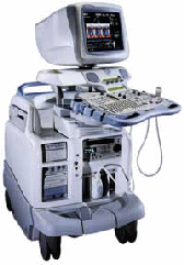 From GE Healthcare.;
'Vivid 7 Dimension, a premier cardiovascular ultrasound system from GE Healthcare, expands on the strength of a powerful imaging platform to offer new, innovative technology of dimensional proportions.'
Device Information and Specification
CONFIGURATION
Multi-frequency, linear, convex, phased, sector
B-mode, C-mode, M-mode (and 2-D), triplex mode, harmonic imaging, color flow mapping, 3D ultrasound display, power Doppler imaging (PDI), color Doppler, pulsed wave Doppler, continuous wave Doppler, tissue velocity imaging (TVI), tissue type imaging (TTI), strain rate imaging (SRI), tissue synchronization imaging (TSI)
IMAGING OPTIONS
CINE review with 5 speed types, bi- andtri-plane imaging with e.g. stress echo and tissue synchronization imaging
STORAGE, CONNECTIVITY, OS
Patient and image archive, HDD, MOD, DVD, USB flash card, DICOM 3.0 Windows-based
DATA PROCESSING
Digital beamformer with 1024 system processing channel technology
H*W*D m (inch.)
1.58 * 0.64 * 0.89 (62 * 25 * 35)
WEIGHT
191 kg (420 lbs.)
POWER CONSUMPTION
less than 2 KVA
•  From GE Healthcare.;
'GE is defining a new age of ultrasound. We call it Volume Ultrasound. GE's Voluson 730 Expert is a powerful system that enables real-time techniques for acquiring, navigating and analyzing volumetric images so that you can make clinical decisions with unprecedented confidence.'
Device Information and Specification
APPLICATIONS
Abdominal, breast, cardiac, musculoskeletal, neonatal, OB/GYN, pediatric, small parts, transcranial, urological, vascular
CONFIGURATION
15' high resolution non-interlaced flat CRT, 4 active probe ports
B-mode, M-mode, coded harmonic imaging (2-D), color flow mode (CFM), power Doppler imaging (PDI), color Doppler, pulsed wave Doppler, high pulse repetition frequency (HPRF) Doppler, tissue harmonic imaging, 3-D power Doppler
IMAGING OPTIONS
CrossXBeam spatial compounding, coded excitation , spatio-temporal image correlation (STIC), B-Flow (simultaneous imaging of tissue and blood flow), strain rate imaging (SRI)
OPTIONAL PACKAGE
STORAGE, CONNECTIVITY, OS
SonoView archiving and data management, network, HDD, DICOM 3.0, CD/DVD, MOD, USB, Windows-based
DATA PROCESSING
Digital beamformer with 512 system processing channel technology
H*W*D m (inch.)
1.43 * 0.69 * 1.02 (56 * 27 * 40)
WEIGHT
136 kg (300 lbs.)
Result Pages : | Share This Page Look Ups |
Medical-Ultrasound-Imaging.com
former US-TIP.com
Member of SoftWays' Medical Imaging Group - MR-TIP • Radiology TIP • Medical-Ultrasound-Imaging
Copyright © 2008 - 2024 SoftWays. All rights reserved.
Terms of Use | Privacy Policy | Advertise With Us
former US-TIP.com
Member of SoftWays' Medical Imaging Group - MR-TIP • Radiology TIP • Medical-Ultrasound-Imaging
Copyright © 2008 - 2024 SoftWays. All rights reserved.
Terms of Use | Privacy Policy | Advertise With Us
[last update: 2023-11-06 01:42:00]