Medical Ultrasound Imaging
Wednesday, 8 May 2024
Info Sheets Out- side     | '3D Ultrasound' p2 Searchterm '3D Ultrasound' found in 23 articles 1 term [ • ] - 18 definitions [• ] - 4 booleans [• ]Result Pages : •
A-mode (Amplitude-mode) ultrasound is a technique used to assess organ dimensions and determine the depth of an organ. While A-mode technology was previously employed in midline echoencephalography for rapid screening of intracranial mass lesions and ophthalmologic scanning, it is now considered obsolete in medical imaging. Nonetheless, the A-mode scan has found applications in early pregnancy assessment (specifically the detection of fetal heartbeats), cephalometry, and placental localization.
When the ultrasound beam encounters an anatomic boundary, the received sound impulse is processed to appear as a vertical reflection of a point. On the display, it looks like spikes of different heights (the amplitude). The intensity of the returning impulse determined the height of the vertical reflection and the time it took for the impulse to make the round trip would determine the space between verticals. The distance between these spikes can be measured accurately by dividing the speed of sound in tissue (1540 m/sec) by half the sound travel time. During an echoencephalography scan, the first A-mode scan is acquired from the right side of the head and captured on film. Subsequently, the probe is positioned at the corresponding point on the left side, and a second exposure is captured on the same film, displaying inverted spikes. The A-mode ultrasound could be used to identify structures normally located in the midline of the brain such as the third ventricle and falx cerebri. The midline structures would be aligned in normal patients but show displacement in patients with mass lesion such as a subdural, epidural, or intracranial hemorrhage. See also 2D Ultrasound, 3D Ultrasound, 4D Ultrasound, Ultrasound Biomicroscopy, A-scan, B-mode and the Infosheet about ultrasound modes. •
An image artifact is any image attribute, which is not present in the original imaged object. An image artifact is sometime the result of an improper operation of the imager, and in other times a consequence of natural processes or properties of the human body. Artifacts in diagnostic ultrasound are a reflection or an echo, which appears on the display and represents the real anatomical structure not correctly. An artifact can be a false, multiple or misleading information introduced by the imaging system or by interaction of ultrasound with the adjacent tissue. Artifacts in ultrasound can be classified as to their source like e.g.:
•
physiologic (motion, different sound velocities, acoustical impedances of tissue);
•
hardware (dimension of the ultrasound beam and the transducer array);
•
Image artifacts can occur in each medical ultrasound. Then an interpretation of the image is complicated and can eliminate the structural information of objects looking for. See also Ultrasound Imaging Procedures. •
A C-scan ultrasound can be displayed in 2D or 3D ultrasound technique. C-scan systems can generate images which are parallel to the surface of the skin (coronal). 2D plane images, usually in gray scale, are recordable at different depths, maintaining high quality subsurface information.
Further Reading: Basics:
News & More:
•  From Hitachi Medical Corporation (HMC);
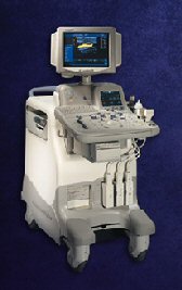
The HI VISION™ 6500 - EUB-6500 high resolution digital ultrasound system offers advanced clinical imaging, enhanced operating efficiency, and remarkable clinical flexibility, all in robust and versatile configuration that simply represents a better clinical solution in a variety of real-world, real-work arenas.
Device Information and Specification
APPLICATIONS
Abdominal, brachytherapy/cryotherapy, breast, cardiac, dedicated biopsy, endoscopic, intraoperative, laparoscopic, musculoskeletal, OB/GYN, pediatric, small parts, urologic, vascular
CONFIGURATION
Compact system
Linear, convex, radial, miniradial/miniprobe, biplane, phased array, echoendoscope longitudinal, echoendoscope radial
Tissue Doppler imaging (TDI), pulsed wave Doppler, continuous wave Doppler, color flow imaging, power Doppler, directional power Doppler, color flow angiography, real-time Doppler measurements, 4 modes of dynamic tissue harmonic imaging (dTHI), wideband pulse inversion imaging (WPI)
IMAGING OPTIONS
3RD generation color artifact suppression
OPTIONAL PACKAGE
3D ultrasound, dual omni-directional M-mode display, steerable CW Doppler, dynamic contrast harmonics imaging, stress echo, Pentax EUS and Fujinon Mini-probe
STORAGE, CONNECTIVITY, OS
Patient and image database management system, HDD, FDD, MOD, CD-ROM, Network, DICOM 3.0, Windows XP
DATA PROCESSING
H*W*D m (inch.)
1.40 x 0.51 x 0.79 (55 x 20 x 31)
WEIGHT
130 kg (286 lbs.)
POWER CONSUMPTION
1.2kVA
ENVIRONMENTAL POLLUTION
4096 btu/hr heat output
•  From GE Healthcare.;
Versatile High Performance System 'The LOGIQ 5 Expert ultrasound system delivers the premium performance advantage of TruScan architecture in a versatile high performance package. Advanced capabilities for outstanding clinical performance are available with the efficiency and productivity needed to meet clinical demands.'
Device Information and Specification
APPLICATIONS
Abdominal, cardiac, musculoskeletal, neonatal, OB/GYN, small parts, transcranial, urologic, vascular
CONFIGURATION
Normal system
B-mode, M-mode, anatomic M-mode creation and adjustment, triplex, 3D ultrasound, pulsed wave Doppler, continuous wave Doppler, power Doppler, color Doppler, spectral Doppler
IMAGING OPTIONS
OPTIONAL PACKAGE
STORAGE, CONNECTIVITY, OS
On-board patient, image and reporting archive, HDD, CD-ROM disk burner included, PCMCIA, USB, Windows-based OS
DATA PROCESSING
H*W*D m (inch.)
1.45 * 0.52 * 0.99 (57 * 20 * 39)
WEIGHT
180 kg (397 lbs.)
POWER CONSUMPTION
less than 1.5 KVA
Result Pages : | Share This Page Look Ups |
Medical-Ultrasound-Imaging.com
former US-TIP.com
Member of SoftWays' Medical Imaging Group - MR-TIP • Radiology TIP • Medical-Ultrasound-Imaging
Copyright © 2008 - 2024 SoftWays. All rights reserved.
Terms of Use | Privacy Policy | Advertise With Us
former US-TIP.com
Member of SoftWays' Medical Imaging Group - MR-TIP • Radiology TIP • Medical-Ultrasound-Imaging
Copyright © 2008 - 2024 SoftWays. All rights reserved.
Terms of Use | Privacy Policy | Advertise With Us
[last update: 2023-11-06 01:42:00]