Medical Ultrasound Imaging
Monday, 13 May 2024
Info Sheets Out- side     | 'Ultrasound Imaging Procedures' p3 Searchterm 'Ultrasound Imaging Procedures' found in 15 articles 1 term [ • ] - 5 definitions [• ] - 9 booleans [• ]Result Pages : •
Common ultrasound supplies that are often used in conjunction with ultrasound imaging:
•
Ultrasound Gel: A water-based gel used as a coupling agent between the transducer and the patient's skin. It helps eliminate air pockets and ensures good sound wave transmission. •
Probe Covers: Disposable covers designed to maintain hygiene and prevent cross-contamination. These covers are placed over the transducer before each examination. •
Cleaning Wipes: Alcohol-based or disinfectant wipes used for cleaning and disinfecting the transducer and other equipment surfaces. Specific cleaning solutions are recommended by the ultrasound equipment manufacturer for thorough cleaning of transducers. •
Gel Warmers: Devices used to warm ultrasound gel, providing patient comfort during the examination. •
Needle Guides: Attachments or brackets that assist in accurate needle placement during ultrasound-guided procedures such as biopsies or injections. •
Positioning Aids: Cushions, wedges, or straps designed to help position patients correctly and comfortably during ultrasound exams. Common ultrasound accessories that are often used in conjunction with ultrasound imaging: •
Transducer Storage Rack: A dedicated rack or holder to store transducers safely when not in use, helping to prevent damage. •
Storage and Archiving Solutions: External hard drives, network storage, or cloud-based systems for long-term storage and backup of ultrasound images and reports. Possibly specialized printers that produce hard copies of ultrasound images for immediate documentation and patient records. •
Power Supply and Transducer Cable Extenders: Extension cables used to increase the length of transducer cables for more flexibility during examinations. Adequate power sources or uninterrupted power supply (UPS) to ensure continuous operation of the ultrasound machine during power outages or fluctuations. •
Reporting Templates and Software: Customizable reporting templates and software solutions that facilitate efficient and standardized reporting of ultrasound findings. •
Phantom Devices: Artificial tissue-like structures or phantoms used for training, calibration, and quality assurance purposes to evaluate image quality and system performance. Consult with ultrasound equipment vendors or professionals in the field to determine the specific accessories and supplies that best suit your imaging needs and specialty. See also Equipment Preparation, Environmental Protection, Portable Ultrasound Machine, Ultrasound Technology, Ultrasound System Performance and Sonographer. • 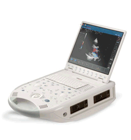 From ESAOTE S.p.A.;
'The MyLab™30CV ultrasound system is an evolutionary step in ultrasound technology. Weighing less than 20 pounds, it is the first compact ultrasound system to deliver premium console performance. And with mobile, portable or stationary configurations, MyLab30CV can adapt to any clinical environment.'
Device Information and Specification
APPLICATIONS
Abdominal, breast, cardiac, OB/GYN, pediatric, pediatric cardiology, small parts, transcranial, vascular
CONFIGURATION
Portable
Linear: 4-10 MHz, convex: 2-5 MHz, phased: 1.6-10 MHz, micro convex: 5-7.5 MHz, endocavity: 5-7.5 MHz, pencil: 2 + 5 MHz
2-D, M-mode, duplex, triplex, color Doppler, pulsed wave Doppler, tissue velocity mapping (TVM), tissue enhancement imaging (TEI™), contrast harmonic imaging, stress echo, tissue velocity mapping for LV motion analysis (TVM), contrast tuned imaging for contrast media procedures (CnTI™), Qontrast™ for myocardium parameters quantification
STORAGE, CONNECTIVITY, OS
Digital patient archive/management, integrated CD/RW, RJ 45 and USB ports, Windows
H*W*D m (inch.)
0.16 * 0.36 * 0.50 (6.2 x 14 x 19.3)
WEIGHT
Less than 11 kg (20 lbs.)
•
The definition of imaging is the visual representation of an object. Medical imaging is a broad term that encompasses various imaging modalities and techniques used in the field of medicine to visualize and study the body's anatomy and physiology. It includes both diagnostic and non-diagnostic imaging procedures, where diagnostic imaging specifically refers to the subset of medical imaging techniques that are primarily focused on diagnosing diseases or conditions. Medical imaging techniques are employed to obtain images or visual representations of the internal organs, tissues, and structures, aiding in the diagnosis, treatment, and monitoring of medical conditions.
The field of medical imaging has significantly evolved since the discovery of X-rays by Konrad Roentgen in 1896. Initially, radiological imaging involved focusing X-rays on the body and capturing the images on a single piece of film within a specialized cassette. Subsequent advancements introduced the use of fluorescent screens and special glasses for real-time visualization of X-ray images. A significant breakthrough came with the application of contrast agents, enhancing image contrast and improving organ visualization. In the 1950s, nuclear medicine studies utilizing gamma cameras demonstrated the uptake of low-level radioactive chemicals in organs, enabling the observation of biological processes in vivo. Currently, positron emission tomography (PET) and single photon emission computed tomography (SPECT) technologies play pivotal roles in clinical research and the diagnosis of biochemical and physiological processes. Additionally, the advent of the x-ray image intensifier in 1955 facilitated the capture and display of x-ray movies. In the 1960s, diagnostic imaging incorporated the principles of sonar, using ultrasonic waves generated by a quartz crystal. These waves, reflecting at the interfaces between different tissues, were received by ultrasound machines and translated into images through computer algorithms and reconstruction software. Ultrasound (ultrasonography) has become an indispensable diagnostic tool across various medical specialties, with immense potential for further advancements such as targeted contrast imaging, real-time 3D or 4D ultrasound, and molecular imaging. The first use of ultrasound contrast agents (USCA) dates back to 1968. Digital imaging techniques were introduced in the 1970s, revolutionizing conventional fluoroscopic image intensifiers. Godfrey Hounsfield's pioneering work led to the development of the first computed tomography (CT) scanner. Digital images are now electronic snapshots represented as grids of dots or pixels. X-ray CT brought about a breakthrough in medical imaging by providing cross-sectional images of the human body with high contrast between different types of soft tissue. These advancements were made possible by analog-to-digital converters and computers. The introduction of multislice spiral CT technology dramatically expanded the clinical applications of CT scans. The first magnetic resonance imaging (MRI) devices were tested on clinical patients in 1980. With technological improvements, such as higher field strength, more open MRI magnets, faster gradient systems, and novel data-acquisition techniques, MRI has emerged as a real-time interactive imaging modality capable of providing detailed structural and functional information of the body. Today, imaging in medicine offers a wide range of modalities, including:
•
X-ray projection imaging;
•
Fluoroscopy;
•
Computed tomography (CT / CAT);
•
Ultrasound imaging (US)
•
Magnetic resonance imaging (MRI), Magnetic source imaging (MSI);
•
Single photon emission computed tomography (SPECT);
•
Positron emission tomography (PET);
•
Mammography.
These imaging modalities have become integral components of modern healthcare. With the rapid advancement of digital imaging, efficient management has become important, leading to the expansion of radiology information systems (RIS) and the adoption of Picture Archiving and Communication Systems (PACS) for digital image archiving. In telemedicine, real-time transmission of all medical image modalities from MRI to X-ray, CT and ultrasound has become the standard. The field of medical imaging continues to evolve, promising further innovations and advancements in the future, ultimately contributing to improved patient care and diagnostics. See also History of Ultrasound Contrast Agents, and History of Ultrasound. Further Reading: News & More:
•
The earliest introduction of vascular ultrasound contrast agents (USCA) was by Gramiak and Shah in 1968, when they injected agitated saline into the ascending aorta and cardiac chambers during echocardiographic to opacify the left heart chamber. Strong echoes were produced within the heart, due to the acoustic mismatch between free air microbubbles in the saline and the surrounding blood.
•
In 1880 the Curie brothers discovered the piezoelectric effect in quartz. Converse piezoelectricity was mathematically deduced from fundamental thermodynamic principles by Lippmann in 1881.
•
In 1917, Paul Langevin (France) and his coworkers developed an underwater sonar system (called hydrophone) that uses the piezoelectric effect to detect submarines through echo location.
•
In 1935, the first RADAR system was produced by the British physicist Robert Watson-Wat. Also about 1935, developments began with the objective to use ultrasonic power therapeutically, utilizing its heating and disruptive effects on living tissues. In 1936, Siemens markets the first ultrasonic therapeutic machine, the Sonostat.
•
Shortly after the World War II, researchers began to explore medical diagnostic capabilities of ultrasound. Karl Theo Dussik (Austria) attempted to locate the cerebral ventricles by measuring the transmission of ultrasound beam through the skull. Other researchers try to use ultrasound to detect gallstones, breast masses, and tumors. These first investigations were performed with A-mode.
•
Shortly after the World War II, researchers in Europe, the United States and Japan began to explore medical diagnostic capabilities of ultrasound. Karl Theo Dussik (Austria) attempted to locate the cerebral ventricles by measuring the transmission of ultrasound beam through the skull. Other researchers, e.g. George Ludwig (United States) tried to use ultrasound to detect gallstones, breast masses, and tumors. This first experimentally investigations were performed with A-mode. Ultrasound pioneers contributed innovations and important discoveries, for example the velocity of sound transmission in animal soft tissues with a mean value of 1540 m/sec (still in use today), and determined values of the optimal scanning frequency of the ultrasound transducer.
•
In the early 50`s the first B-mode images were obtained. Images were static, without gray-scale information in simple black and white and compound technique. Carl Hellmuth Hertz and Inge Edler (Sweden) made in 1953 the first scan of heart activity. Ian Donald and Colleagues (Scotland) were specialized on obstetric and gynecologic ultrasound research. By continuous development it was possible to study pregnancy and diagnose possible complications.
•
After about 1960 two-dimensional compound procedures were developed. The applications in obstetric and gynecologic ultrasound boomed worldwide from the mid 60's with both, A-scan and B-scan equipment. In the late 60's B-mode ultrasonography replaced A-mode in wide parts.
•
In the 70's gray scale imaging became available and with progress of computer technique ultrasonic imaging gets better and faster.
•
•
In the 90's, high resolution scanners with digital beamforming, high transducer frequencies, multi-channel focus and broad-band transducer technology became state of the art. Optimized tissue contrast and improved diagnostic accuracy lead to an important role in breast imaging and cancer detection. Color Doppler and Duplex became available and sensitivity for low flow was continuously improved.
•
Actually, machines with advanced ultrasound system performance are equipped with realtime compound imaging, tissue harmonic imaging, contrast harmonic imaging, vascular assessment, matrix array transducers, pulse inversion imaging, 3D and 4D ultrasound with panoramic view.
• The field of medical imaging offers numerous career opportunities, and one profession is that of a sonographer. Sonographers play a critical role in healthcare by utilizing ultrasound technology to create images of the body's internal structures. •
Becoming a Sonographer: The educational and professional requirements for sonographers can vary from country to country. The duration of these programs can range from one to four years, depending on the country and level of qualification. The typical path in the United States begins with obtaining a post-secondary education in diagnostic medical sonography from an accredited program. These programs usually result in an associate's or bachelor's degree. Coursework typically covers anatomy, physiology, medical ethics, ultrasound physics, and specialized sonography techniques. Additionally, students gain practical experience through clinical internships in healthcare facilities. After completing their education, aspiring sonographers can choose to obtain professional certification through organizations such as the American Registry for Diagnostic Medical Sonography (ARDMS) or the American Registry of Radiologic Technologists (ARRT). Certification often requires passing examinations that assess knowledge and competency in specific areas of sonography. Many countries also have certification or registration requirements for sonographers. These certifications are typically obtained through professional bodies or organizations specific to each country. Examples include the Canadian Association of Registered Diagnostic Ultrasound Professionals (CARDUP) in Canada, the Australian Sonographers Accreditation Registry (ASAR) in Australia, and the Society and College of Radiographers (SCoR) in the United Kingdom. •
Job Description: Sonographers are skilled professionals who operate ultrasound machines and perform sonograms on patients. They work closely with physicians and other healthcare professionals to provide accurate and high-quality diagnostic images. Using sound waves, sonographers capture images of organs, tissues, and blood flow patterns, which are then used by medical practitioners to diagnose and monitor various medical conditions. Sonographers must have a comprehensive understanding of anatomy, physiology, and sonographic techniques to optimize image quality. They interact directly with patients, explaining procedures, addressing concerns, and ensuring patient comfort throughout the scanning process. Documentation of findings and communication with the medical team are also essential responsibilities. Some aspect of the job can be demanding, while sonographers often spend long hours on their feet, positioning and maneuvering patients during scans. Dealing with patients who are in pain, anxious, or difficult to scan requires empathy, patience, and excellent interpersonal skills. Sonographers often work in fast-paced environments, juggling multiple patients and procedures throughout the day. Effective time management is essential to ensure that scans are performed efficiently without compromising quality. Adhering to schedules and meeting the demands of the healthcare facility can add to the workload and stress levels. •
Salary Outlook: The salary of a sonographer can vary, based on factors such as experience, specialization, geographic location, and work setting. According to the U.S. Bureau of Labor Statistics, as of May 2021, the median annual wage for diagnostic medical sonographers was $77,740. Sonographers working in specialized hospitals, outpatient care centers, and diagnostic imaging centers tend to earn higher salaries compared to those in physician offices or government facilities. The salary prospects for sonographers outside the United States can vary significantly based on factors such as the country's economic conditions, healthcare system, demand for sonographers, and cost of living. •
Future Outlook: The future outlook for sonographers appears highly favorable. The demand for ultrasound imaging continues to grow due to advancements in medical technology and an aging population. This increasing demand for sonographers is expected to result in good job prospects and potential career advancement opportunities. Monitoring job markets, understanding regulatory requirements, and networking with professionals in international healthcare communities can provide valuable insights into future opportunities. See also Handheld Ultrasound, Ultrasound Machine, Sonography, Portable Ultrasound Machine, Ultrasound Accessories and Supplies, Environmental Protection and Ultrasound Technology. Result Pages : | Share This Page Look Ups |
Medical-Ultrasound-Imaging.com
former US-TIP.com
Member of SoftWays' Medical Imaging Group - MR-TIP • Radiology TIP • Medical-Ultrasound-Imaging
Copyright © 2008 - 2024 SoftWays. All rights reserved.
Terms of Use | Privacy Policy | Advertise With Us
former US-TIP.com
Member of SoftWays' Medical Imaging Group - MR-TIP • Radiology TIP • Medical-Ultrasound-Imaging
Copyright © 2008 - 2024 SoftWays. All rights reserved.
Terms of Use | Privacy Policy | Advertise With Us
[last update: 2023-11-06 01:42:00]