Medical Ultrasound Imaging
Monday, 20 May 2024
Info Sheets Out- side     | 'Endoscopic Ultrasound' p2 Searchterm 'Endoscopic Ultrasound' found in 11 articles 1 term [ • ] - 6 definitions [• ] - 4 booleans [• ]Result Pages : •
(TRUS) Transrectal sonography (also called transrectal ultrasonography, transrectal echography (TRE), endorectal ultrasound (ERUS or EUS)) is an ultrasound procedure used to examine the prostate gland, the rectum or bladder. A small, lubricated transducer placed into the rectum releases sound waves, which create echoes as they enter the region of interest. A computer creates a picture called a sonogram. TRUS is commonly used for guidance during a prostate needle biopsy and may be used to deliver brachytherapy and monitor cancer treatment. Transrectal ultrasonography detects enlargement, tumors and other abnormalities of the prostate, rectal polyps, rectal cancer, perianal infection, and sphincter muscle injuries. TRUS is also performed on male patients with infertility to view the prostate and surrounding structures and on patients with suspected bladder conditions or disease to view the bladder. See also Transurethral Sonography, Endoscopic Ultrasound, Pelvic Ultrasound, Rectal Probe, Biplane Probe, Endocavitary Echography and High Intensity Focused Ultrasound. Further Reading: News & More:
•
Ultrasound imaging procedures are widely used in medicine. It is possible to perform diagnostic or therapeutic procedures with the guidance of ultrasonography (interventional ultrasound biopsies or drainage of fluid collections). Sonography or ultrasound scanning involves the application of an ultrasound transducer used to transmit high frequency sound waves, which bounce off internal structures to produce an image that can be displayed and recorded.
Ultrasound imaging procedures include for example: Further Reading: News & More:
•
(TEE) Transesophageal echocardiography provides a superior view of cardiac anatomy compared with transthoracic echocardiography. TEE is performed by the introduction of a probe attached to a fiberoptic endoscope into the esophagus. Caused by the position close to the heart e.g., clot finding and the view of the mitral valve are improved. Indications:
•
aortic atherosclerotic disease;
•
aortic dissection;
•
artificial mitral valves;
•
clots inside the left atrium;
•
cardiac infections;
•
masses or clots in the heart.
The piezoelectric crystal creating the acoustic power is mounted on the gastroscope that must be swallowed by the patient. This endoscopic transducer is miniaturized to approximately the size of a fingernail. Usually the probe is in place for an average of 15 minutes, to numb the surface a topical anesthetic is sprayed into the throat, in addition a conscious sedation is recommended. See also Myocardial Contrast Echocardiography, Stress Echocardiogram, M-Mode Echocardiography, Contrast Enhanced Ultrasound and Vascular Ultrasound Contrast Agents. • 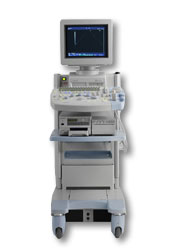 From Hitachi Medical Corporation (HMC), sales, marketing and service in the US by Hitachi Medical Systems America Inc.
The HI VISION™ 5500 - EUB-5500 fully digital ultrasound system delivers the latest generation of signal processing technology, sophisticated transducer design, and a host of features and options for advanced imaging capabilities across a wide range of clinical situations. This system is compatible with all Pentax ultrasound endoscopes.
Device Information and Specification
APPLICATIONS
Abdominal, brachytherapy/cryotherapy, breast, cardiac, dedicated biopsy, endoscopic, intraoperative, laparoscopic, musculoskeletal, OB/GYN, pediatric, small parts, urologic, vascular
CONFIGURATION
Compact system
Five frequency (except mini-probes)
Linear, convex, radial, miniradial/miniprobe, biplane, phased array, echoendoscope longitudinal, echoendoscope radial
3 modes of dynamic tissue harmonic imaging (dTHI), pulsed wave Doppler, continuous wave Doppler, color flow imaging, power Doppler, directional power Doppler, color flow angiography, real-time Doppler measurements
IMAGING OPTIONS
3RD generation color artifact suppression
OPTIONAL PACKAGE
STORAGE, CONNECTIVITY, OS
Patient and image database management system, HDD, FDD, MOD, CD-ROM, Network, DICOM 3.0, Windows XP
DATA PROCESSING
H*W*D m (inch.)
1.40 x 0.51 x 0.79 (55 x 20 x 31)
WEIGHT
130 kg (286 lbs.)
POWER CONSUMPTION
1.2kVA
ENVIRONMENTAL IMPACT
4096 btu/hr heat output
•  From Hitachi Medical Corporation (HMC), sales, marketing and service in the US by Hitachi Medical Systems America Inc.;
Powerful, flexible, and fast, the HI VISION™ 8500 - EUB-8500 diagnostic ultrasound scanner combines leading edge technologies with user-oriented operation for exceptional imaging and functionality. Available exclusively on the 8500, SonoElastography provides a new perspective on the physical properties of tumors and masses by determining and displaying the relative stiffness of tissue. Device Information and Specification
APPLICATIONS
Abdominal, brachytherapy/cryotherapy, breast, cardiac, dedicated biopsy, endoscopic, intraoperative, laparoscopic, musculoskeletal, OB/GYN, pediatric, small parts, urologic, vascular
CONFIGURATION
Compact system
RANGE OF PROBE TYPE
Linear, convex, radial, biplane, phased array, echoendoscope longitudinal, echoendoscope radial
PROBE FREQUENCIES
Linear: 5.0-13 MHz, convex: 2.5-7.5 MHz, phased:
2.0-7.5 MHz, sector: 2.0-7.5 MHz
4 Modes of dynamic tissue harmonic imaging (dTHI), pulsed wave Doppler, continuous wave Doppler, color flow imaging, power Doppler, directional power Doppler, color flow angiography, real-time Doppler measurements, quantitative tissue Doppler
IMAGING OPTIONS
HI COMPOUND imaging,
HI RES adaptive imaging, wideband pulse inversion imaging (WPI), Raw Data Freeze
OPTIONAL PACKAGE
IMAGING ENHANCEMENTS
3RD generation color artifact suppression
STORAGE, CONNECTIVITY, OS
Patient and image database management system, HDD, FDD, MOD, CD-ROM, Network, DICOM 3.0, Windows XP
DATA PROCESSING
Octal beam processing, 12 bit Gigasampling A/D for precise signal reproduction
H*W*D m (inch.)
1.50 * 0.56 * 1.02 (59 x 22 x 40)
WEIGHT
159 kg (351 lbs.)
POWER CONSUMPTION
1.5kVA
Result Pages : | Share This Page Look Ups |
Medical-Ultrasound-Imaging.com
former US-TIP.com
Member of SoftWays' Medical Imaging Group - MR-TIP • Radiology TIP • Medical-Ultrasound-Imaging
Copyright © 2008 - 2024 SoftWays. All rights reserved.
Terms of Use | Privacy Policy | Advertise With Us
former US-TIP.com
Member of SoftWays' Medical Imaging Group - MR-TIP • Radiology TIP • Medical-Ultrasound-Imaging
Copyright © 2008 - 2024 SoftWays. All rights reserved.
Terms of Use | Privacy Policy | Advertise With Us
[last update: 2023-11-06 01:42:00]