Medical Ultrasound Imaging
Wednesday, 8 May 2024
Info Sheets Out- side     | 'Image Quality' p3 Searchterm 'Image Quality' found in 40 articles 1 term [ • ] - 39 definitions [• ] Result Pages : •  From SIUI Inc.;
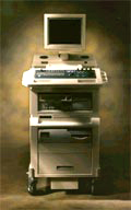
'Dedicated to ultrasound industry, Shantou Institute of Ultrasonic Instruments, Inc. (SIUI) has launched Apogee 3500, the Digital Color Doppler Ultrasound Imaging System. With latest imaging technologies, high-definition image quality and excellent practical functions, the Apogee 3500 offers optimal solutions for clinical ultrasonic examination.' 'The Apogee 3500 is available with many high-density, super broadband and multi-frequency probes, such as convex, micro-convex, linear, vaginal, rectal and phased array probes, which are widely applied for different clinical diagnoses, including abdomen (liver, kidney, gall-bladder, pancreas), gynecology (uterus, ovary), obstetrics (early pregnancy, basic OB, complete OB, multi gestation, fetal echo), cardiology (adult and pediatric cardiology), small parts (thyroid, galactophore, testicles, neonate), peripheral vascular and prostate.'
Device Information and Specification
CONFIGURATION
Normal system, color - gray scale(256)
Linear, convex and phased array
PROBES STANDARD
2.0 MHz ~ 12.0 MHz, broad band, tri-frequency
B-mode (B, 2B, 4B), M-mode, B/M-mode, real-time compound imaging, panoramic imaging, trapezoidal imaging (linear probes), spectrum Doppler (PWD and CWD), color Doppler flow imaging (CDFI), color power angio (CPA), tissue harmonic imaging (THI)
IMAGING OPTIONS
Real-time ZOOM, zoom rate and position selectable
OPTIONAL PACKAGE
H*W*D m
1.29 * 0.52 * 0.75
WEIGHT
110 kg
POWER REQUIREMENT
AC 220V/110V, 50Hz/60Hz
POWER CONSUMPTION
0.6 KVA
•  From SIUI Inc.;
'The Apogee 800Plus ultrasound system offers a new level of high quality in image. With broad clinical application, the system delivers ease of use for cardiac and vascular exams.' 'Due to the exceptional image quality, the sensitivity of color Doppler and many other advanced features, the Apogee 800Plus offers complete diagnosis capability for your entire patient population, including abdominal, obstetrics, gynecology, adult cardiology, pediatric cardiology, peripheral vascular, small parts, breast, prostate, transcranial, etc.'
Device Information and Specification
APPLICATIONS
Linear, curved, phased and symmetric phased array
2D, M-mode, Pulsed wave Doppler, Color Power Angio Imaging, Color 2D, Color M-mode, High PRF Doppler
H*W*D m
1.46 * 0.69 * 0.87
WEIGHT
140 kg (without peripherals)
POWER REQUIREMENT
115Vac, (V~), @ 50 to 60 Hz, 1.2kVA
230Vac, (V~), @ 50 to 60 Hz, 1.2kVA POWER CONSUMPTION
800 Watts (with optional OEM'S: 1200 Watts max)
•
The wider the ultrasound beam, the more severe the problem with volume averaging and the beam-width artifact, to avoid this, the ultrasound beam can be shaped with lenses.
Different possibilities to focus the beam:
•
Mechanical focusing is performed by placing an acoustic lens on the surface of the transducer or using a transducer with a concave face.
•
Electronic focusing uses multiple phased array (annular or linear) elements, sequentially fired to focus the beam.
Conventional multi-element transducers are electronically focused in order to minimize beam width. This transducer type can be focused electronically only along the long axis of the probe where there are multiple elements, along the short axis (elevation axis) are conventional transducers only one element wide. Electronic focusing in any axis requires multiple transducer elements arrayed along that axis. Short axis focusing of conventional multi-element transducers requires an acoustic lens which has a fixed focal length. For operation at frequencies at or even above 10 MHz, quantization noise reduces contrast resolution. Digital beamforming gives better control over time delay quantization errors. In digital beamformers the delay accuracy is improved, thus allowing higher frequency operation. In analog beamformers, delay accuracy is in the order of 20 ns. Phased beamformers are suitable to handle linear phased arrays and are used for sector formats such as required in cardiography to improve image quality. Beamforming in ultrasound instruments for medical imaging uses analog delay lines. The signal from each individual element is delayed in order to steer the beam in the desired direction and focuses the beam. The receive beamformer tracks the depth and focuses the receive beam as the depth increases for each transmitted pulse. The receive aperture increase with depth. The lateral resolution is constant with depth, and decreases the sensitivity to aberrations in the imaged tissue. A requirement for dynamic control of the used elements is given. Since often a weighting function (apodization) is used for side lobe reduction, the element weights also have to be dynamically updated with depth. See also Huygens Principle. •
Increasing the frequency of the transmitted power improves the image quality of ultrasound, but the improvement in resolution results in a decreased signal to noise ratio (SNR). Higher acoustic power levels can prevent the loss in SNR, but among other reasons, ultrasound regulations limit this to avoid heating or cavitation. Coded excitation increase the signal to noise ratio without the loss of resolution by using coded waveforms. Coded excitation allows transmitting a long wide-band pulse with more acoustic power and high penetration of the sound beam. •
Compound B-mode imaging takes different forms and refers to different methods of creating the ultrasound image. Real-time compound ultrasound improves the image quality of B-mode scanning by combining ultrasound information obtained from multiple angles. The used averaging process of compound B-mode reduces artifacts and improves the representation of true image data. B-mode images and Doppler mode images (see also Duplex) can be compounded on the display to improve the visualization of the anatomical relationships between vessels and the surrounding tissues. Further Reading: News & More:
Result Pages : | Share This Page Look Ups |
Medical-Ultrasound-Imaging.com
former US-TIP.com
Member of SoftWays' Medical Imaging Group - MR-TIP • Radiology TIP • Medical-Ultrasound-Imaging
Copyright © 2008 - 2024 SoftWays. All rights reserved.
Terms of Use | Privacy Policy | Advertise With Us
former US-TIP.com
Member of SoftWays' Medical Imaging Group - MR-TIP • Radiology TIP • Medical-Ultrasound-Imaging
Copyright © 2008 - 2024 SoftWays. All rights reserved.
Terms of Use | Privacy Policy | Advertise With Us
[last update: 2023-11-06 01:42:00]