Medical Ultrasound Imaging
Sunday, 19 May 2024
Info Sheets Out- side     | 'Pulsed Wave Doppler' p3 Searchterm 'Pulsed Wave Doppler' found in 27 articles 1 term [ • ] - 18 definitions [• ] - 8 booleans [• ]Result Pages : • 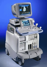 From GE Healthcare.;
'The System of Choice for General Imaging Imagine a leading-edge ultrasound system so versatile that it can meet the demands of virtually any clinical setting. With the LOGIQ® 9, you'll have a high-performance system capable of multi-dimensional imaging for a full range of clinical applications - from abdominal to breast to vascular imaging. And an ergonomic design that improves scanning comfort and clinical work flow. Now, imagine what LOGIQ® 9 could do for you and your patients.'
Device Information and Specification
APPLICATIONS
Abdominal, cardiac, breast, intraoperative, musculoskeletal, neonatal, OB/GYN, orthopedic, pediatric, small parts, transcranial, urologic, vascular
CONFIGURATION
17' high resolution non-interlaced flat CRT, 4 active probe ports
B-mode, M-mode, coded harmonic imaging, color flow mode (CFM), power Doppler imaging (PDI), PW-HPRF, CW Doppler, color Doppler, pulsed wave Doppler, tissue harmonic imaging
IMAGING OPTIONS
CrossXBeam spatial compounding, coded ultrasound acquisition), speckle reduction imaging (SRI), TruScan technology store raw data, real-time 4D ultrasound, Tru 3D ultrasound
STORAGE, CONNECTIVITY, OS
Patient and image archive, HDD, DICOM 3.0, CD/DVD, MOD, PCMCIA, USB, Windows-based
DATA PROCESSING
Digital beamformer with 1024 system processing channel technology
H*W*D m (inch.)
1.62 * 0.61 * 0.99 (64 * 24 * 39)
WEIGHT
202 kg (408 lb.)
POWER CONSUMPTION
less than 2 KVA
•  From ESAOTE S.p.A.;
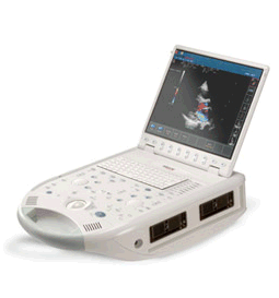
'The MyLab™30CV ultrasound system is an evolutionary step in ultrasound technology. Weighing less than 20 pounds, it is the first compact ultrasound system to deliver premium console performance. And with mobile, portable or stationary configurations, MyLab30CV can adapt to any clinical environment.'
Device Information and Specification
APPLICATIONS
Abdominal, breast, cardiac, OB/GYN, pediatric, pediatric cardiology, small parts, transcranial, vascular
CONFIGURATION
Portable
Linear: 4-10 MHz, convex: 2-5 MHz, phased: 1.6-10 MHz, micro convex: 5-7.5 MHz, endocavity: 5-7.5 MHz, pencil: 2 + 5 MHz
2-D, M-mode, duplex, triplex, color Doppler, pulsed wave Doppler, tissue velocity mapping (TVM), tissue enhancement imaging (TEI™), contrast harmonic imaging, stress echo, tissue velocity mapping for LV motion analysis (TVM), contrast tuned imaging for contrast media procedures (CnTI™), Qontrast™ for myocardium parameters quantification
STORAGE, CONNECTIVITY, OS
Digital patient archive/management, integrated CD/RW, RJ 45 and USB ports, Windows
H*W*D m (inch.)
0.16 * 0.36 * 0.50 (6.2 x 14 x 19.3)
WEIGHT
Less than 11 kg (20 lbs.)
•  From Siemens Medical Systems;
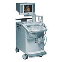
'This extremely flexible system supports targeted applications for OB/GYN, Radiology, Internal Medicine, Urology, and Prostate Brachytherapy. The large transducer selection, most with high-frequency capabilities, provides the right tool for general imaging, radiology, and internal medicine applications.'
Device Information and Specification
CLINICAL APPLICATION
OB/GYN, cerebrovascular, orthopedics, urology, peripheral vascular, pediatrics, small parts, surgery, abdominal, musculoskeletal
CONFIGURATION
Mobile, compact
DISPLAY MODES
Broadband, high-fidelity, multi-frequency, linear and curved
PROBE PORTS
Two - four
256
MEASUREMENT/CALCULATION FUNCTIONS
OB/GYN measurements and report package, off-line analysis
OPTIONAL PACKAGE
IMAGE PROCESSING
•
(TCCS) Transcranial color coded sonography is a combination of B-mode and pulsed wave Doppler. TCCS is used to study morphological and functional assessment of the circle of Willis, intracranial hemodynamics caused by extracranial artery stenosis, collateral flow and the vascular supply of intracranial lesion. Color imaging of the intracranial vessels allows placing the spectral Doppler volume correctly. This modality has encouraged the widespread use. Contrast enhanced TCCS analysis of cerebral arteriovenous transit time (cTT) is used as a measure of cerebral microcirculation. The windows that are used for transcranial Doppler examinations include regions where the skull bones are relatively thin or where naturally occurring gaps allow proper penetration of the sound beam. See also A-Mode, Cranial Bone Thermal Index, Transcranial Doppler and Transcranial Window. •
Ultrasound imaging is excellent for diagnosing cysts and other fluids in soft tissue. For ultrasound imaging or ultrasonography, different modes are used to examine the arterial/venous system, heart, pancreas, urinary system, ovaries, spinal cord, joints and more. Power levels, frequencies used, amplification, and beamforming determine the clarity of the image. These things are controlled by the sonographer, interacting with the properties of the ultrasound machine. Various imaging modes:
•
•
•
•
•
Result Pages : | Share This Page Look Ups |
Medical-Ultrasound-Imaging.com
former US-TIP.com
Member of SoftWays' Medical Imaging Group - MR-TIP • Radiology TIP • Medical-Ultrasound-Imaging
Copyright © 2008 - 2024 SoftWays. All rights reserved.
Terms of Use | Privacy Policy | Advertise With Us
former US-TIP.com
Member of SoftWays' Medical Imaging Group - MR-TIP • Radiology TIP • Medical-Ultrasound-Imaging
Copyright © 2008 - 2024 SoftWays. All rights reserved.
Terms of Use | Privacy Policy | Advertise With Us
[last update: 2023-11-06 01:42:00]