Medical Ultrasound Imaging
Friday, 10 May 2024
Info Sheets Out- side     | 'Breast Ultrasound' p3 Searchterm 'Breast Ultrasound' found in 19 articles 1 term [ • ] - 18 booleans [• ]Result Pages : •
(US) Also called echography, sonography, ultrasonography, echotomography, ultrasonic tomography. Diagnostic imaging plays a vital role in modern healthcare, allowing medical professionals to visualize internal structures of the body and assist in the diagnosis and treatment of various conditions. Two terms that are commonly used interchangeably but possess distinct meanings in the field of medical imaging are 'ultrasound' and 'sonography.' Ultrasound is the imaging technique that utilizes sound waves to create real-time images, while sonography encompasses the entire process of performing ultrasound examinations and interpreting the obtained images. Ultrasonography is a synonymous term for sonography, emphasizing the use of ultrasound technology in diagnostic imaging. A sonogram, on the other hand, refers to the resulting image produced during an ultrasound examination. Ultrasonic waves, generated by a quartz crystal, cause mechanical perturbation of an elastic medium, resulting in rarefaction and compression of the medium particles. These waves are reflected at the interfaces between different tissues due to differences in their mechanical properties. The transmission and reflection of these high-frequency waves are displayed with different types of ultrasound modes. By utilizing the speed of wave propagation in tissues, the time of reflection information can be converted into distance of reflection information. The use of higher frequencies in medical ultrasound imaging yields better image resolution. However, higher frequencies also lead to increased absorption of the sound beam by the medium, limiting its penetration depth. For instance, higher frequencies (e.g., 7.5 MHz) are employed to provide detailed imaging of superficial organs like the thyroid gland and breast, while lower frequencies (e.g., 3.5 MHz) are used for abdominal examinations. Ultrasound in medical imaging offers several advantages including:
•
noninvasiveness;
•
safety with no potential risks;
•
widespread availability and relatively low cost.
Diagnostic ultrasound imaging is generally considered safe, with no adverse effects. As medical ultrasound is extensively used in pregnancy and pediatric imaging, it is crucial for practitioners to ensure its safe usage. Ultrasound can cause mechanical and thermal effects in tissue, which are amplified with increased output power. Consequently, guidelines for the safe use of ultrasound have been issued to address the growing use of color flow imaging, pulsed spectral Doppler, and higher demands on B-mode imaging. Furthermore, recent ultrasound safety regulations have shifted more responsibility to the operator to ensure the safe use of ultrasound. See also Skinline, Pregnancy Ultrasound, Obstetric and Gynecologic Ultrasound, Musculoskeletal and Joint Ultrasound, Ultrasound Elastography and Prostate Ultrasound. Further Reading: Basics: News & More:
• 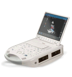 From ESAOTE S.p.A.;
'The MyLab™30CV ultrasound system is an evolutionary step in ultrasound technology. Weighing less than 20 pounds, it is the first compact ultrasound system to deliver premium console performance. And with mobile, portable or stationary configurations, MyLab30CV can adapt to any clinical environment.'
Device Information and Specification
APPLICATIONS
Abdominal, breast, cardiac, OB/GYN, pediatric, pediatric cardiology, small parts, transcranial, vascular
CONFIGURATION
Portable
Linear: 4-10 MHz, convex: 2-5 MHz, phased: 1.6-10 MHz, micro convex: 5-7.5 MHz, endocavity: 5-7.5 MHz, pencil: 2 + 5 MHz
2-D, M-mode, duplex, triplex, color Doppler, pulsed wave Doppler, tissue velocity mapping (TVM), tissue enhancement imaging (TEI™), contrast harmonic imaging, stress echo, tissue velocity mapping for LV motion analysis (TVM), contrast tuned imaging for contrast media procedures (CnTI™), Qontrast™ for myocardium parameters quantification
STORAGE, CONNECTIVITY, OS
Digital patient archive/management, integrated CD/RW, RJ 45 and USB ports, Windows
H*W*D m (inch.)
0.16 * 0.36 * 0.50 (6.2 x 14 x 19.3)
WEIGHT
Less than 11 kg (20 lbs.)
• 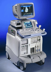 From GE Healthcare.;
'The System of Choice for General Imaging Imagine a leading-edge ultrasound system so versatile that it can meet the demands of virtually any clinical setting. With the LOGIQ® 9, you'll have a high-performance system capable of multi-dimensional imaging for a full range of clinical applications - from abdominal to breast to vascular imaging. And an ergonomic design that improves scanning comfort and clinical work flow. Now, imagine what LOGIQ® 9 could do for you and your patients.'
Device Information and Specification
APPLICATIONS
Abdominal, cardiac, breast, intraoperative, musculoskeletal, neonatal, OB/GYN, orthopedic, pediatric, small parts, transcranial, urologic, vascular
CONFIGURATION
17' high resolution non-interlaced flat CRT, 4 active probe ports
B-mode, M-mode, coded harmonic imaging, color flow mode (CFM), power Doppler imaging (PDI), PW-HPRF, CW Doppler, color Doppler, pulsed wave Doppler, tissue harmonic imaging
IMAGING OPTIONS
CrossXBeam spatial compounding, coded ultrasound acquisition), speckle reduction imaging (SRI), TruScan technology store raw data, real-time 4D ultrasound, Tru 3D ultrasound
STORAGE, CONNECTIVITY, OS
Patient and image archive, HDD, DICOM 3.0, CD/DVD, MOD, PCMCIA, USB, Windows-based
DATA PROCESSING
Digital beamformer with 1024 system processing channel technology
H*W*D m (inch.)
1.62 * 0.61 * 0.99 (64 * 24 * 39)
WEIGHT
202 kg (408 lb.)
POWER CONSUMPTION
less than 2 KVA
• 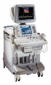 From GE Healthcare.;
'The System of Choice for Shared Service. The LOGIQ® 7 system provides a full range of clinical applications including abdominal, small parts, surgery, vascular and cardiac imaging and the power of GE's patented TruScan architecture. Just imagine an ultrasound system so versatile and reliable that it can meet the demands of virtually any clinical setting. And an ergonomic design that improves scanning comfort and clinical work flow.'
Device Information and Specification
APPLICATIONS
Abdominal, cardiac, breast, intraoperative, musculoskeletal, neonatal, OB/GYN, orthopedic, pediatric, small parts, transcranial, urologic, vascular
CONFIGURATION
17' high resolution non-interlaced flat CRT, 4 active probe ports
B-mode, M-mode, coded harmonic imaging, color flow mode (CFM), power Doppler imaging (PDI), color Doppler, pulsed wave Doppler, tissue harmonic imaging
IMAGING OPTIONS
CrossXBeam spatial compounding, coded ultrasound acquisition),speckle reduction imaging (SRI), TruScan technology store raw data, CINE review with 4 speed types
OPTIONAL PACKAGE
Transesophageal scanning, stress echo, tissue velocity imaging (TVI), tissue velocity Doppler (TVD), contrast harmonic imaging
STORAGE, CONNECTIVITY, OS
Patient and image archive, HDD, DICOM 3.0, CD/DVD, MOD, Windows-based
DATA PROCESSING
Digital beamformer with 1024 system processing channel technology
H*W*D m (inch.)
1.62 * 0.61 * 0.99 (64 * 24 * 39)
WEIGHT
246 kg (498 lbs.)
POWER CONSUMPTION
less than 1.5 KVA
• 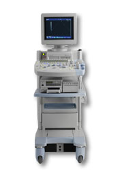 From Hitachi Medical Corporation (HMC), sales, marketing and service in the US by Hitachi Medical Systems America Inc.
The HI VISION™ 5500 - EUB-5500 fully digital ultrasound system delivers the latest generation of signal processing technology, sophisticated transducer design, and a host of features and options for advanced imaging capabilities across a wide range of clinical situations. This system is compatible with all Pentax ultrasound endoscopes.
Device Information and Specification
APPLICATIONS
Abdominal, brachytherapy/cryotherapy, breast, cardiac, dedicated biopsy, endoscopic, intraoperative, laparoscopic, musculoskeletal, OB/GYN, pediatric, small parts, urologic, vascular
CONFIGURATION
Compact system
Five frequency (except mini-probes)
Linear, convex, radial, miniradial/miniprobe, biplane, phased array, echoendoscope longitudinal, echoendoscope radial
3 modes of dynamic tissue harmonic imaging (dTHI), pulsed wave Doppler, continuous wave Doppler, color flow imaging, power Doppler, directional power Doppler, color flow angiography, real-time Doppler measurements
IMAGING OPTIONS
3RD generation color artifact suppression
OPTIONAL PACKAGE
STORAGE, CONNECTIVITY, OS
Patient and image database management system, HDD, FDD, MOD, CD-ROM, Network, DICOM 3.0, Windows XP
DATA PROCESSING
H*W*D m (inch.)
1.40 x 0.51 x 0.79 (55 x 20 x 31)
WEIGHT
130 kg (286 lbs.)
POWER CONSUMPTION
1.2kVA
ENVIRONMENTAL IMPACT
4096 btu/hr heat output
Result Pages : | Share This Page Look Ups |
Medical-Ultrasound-Imaging.com
former US-TIP.com
Member of SoftWays' Medical Imaging Group - MR-TIP • Radiology TIP • Medical-Ultrasound-Imaging
Copyright © 2008 - 2024 SoftWays. All rights reserved.
Terms of Use | Privacy Policy | Advertise With Us
former US-TIP.com
Member of SoftWays' Medical Imaging Group - MR-TIP • Radiology TIP • Medical-Ultrasound-Imaging
Copyright © 2008 - 2024 SoftWays. All rights reserved.
Terms of Use | Privacy Policy | Advertise With Us
[last update: 2023-11-06 01:42:00]