Medical Ultrasound Imaging
Sunday, 19 May 2024
Info Sheets Out- side     | 'Pediatric Ultrasound' p2 Searchterm 'Pediatric Ultrasound' found in 20 articles 1 term [ • ] - 6 definitions [• ] - 13 booleans [• ]Result Pages : •
Sonography [aka: ultrasonography] is a term that encompasses the entire process of performing ultrasound examinations and interpreting the obtained images. Sonography involves the skilled application of ultrasound technology by trained professionals known as sonographers or ultrasound technologists. These specialists operate the ultrasound equipment, manipulate the transducer, and acquire the necessary pictures for diagnostic imaging purposes. Sonography requires in-depth knowledge of anatomy, physiology, and pathology to accurately interpret the ultrasound images and provide valuable information to the treating physician. Sonography uses equipment that generates high frequency sound waves to produce images from muscles, soft tissues, fluid collections, and vascular structures of the human body. Obstetric sonography is commonly used during pregnancy. Sonography visualizes anatomy, function, and pathology of for example gallbladder, kidneys, pancreas, spleen, liver, uterus, ovaries, urinary bladder, eye, thyroid, breast, aorta, veins and arteries in the extremities, carotid arteries in the neck, as well as the heart. A typical medical ultrasound machine, usually a real-time scanner, operates in the frequency range of 2 to 13 megahertz. See also Musculoskeletal and Joint Ultrasound, Pediatric Ultrasound, Cerebrovascular Ultrasonography and Contrast Enhanced Ultrasound. Further Reading: Basics:
News & More:
•
Urologic ultrasound includes the examination of the kidneys, renal vessels, urinary tract, bladder, prostate, and scrotum. Usual gray scale ultrasound equipment and standard probes are sufficient to examine the kidney parenchyma and renal pelvis, the urinary tract and bladder. Doppler ultrasound is a useful adjunct to kidney ultrasound. High ultrasound system performance is desirable to show the arterial system, because advanced power Doppler is significantly more sensitive to blood flow than standard color Doppler. Transurethral sonography may be used to examine the bladder and urethra. Transrectal sonography is used to scan and treat the prostate e.g., with brachytherapy or high intensity focused ultrasound. Very small probes are used for these applications. Reflux sonography is especially used in pediatric ultrasound. See also Ultrasound Imaging Procedures, Ultrasound Picture, Ultrasound Imaging Modes, Lithotripsy, Thermotherapy, Brachytherapy and Ultrasound Therapy. Further Reading: Basics:
News & More:
• 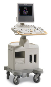 From Philips Medical Systems;
Introduced in June 2005, 'one of the less expensive and more dedicated' ultrasound systems.
Device Information and Specification
CONFIGURATION
LCD monitor
Broadband, convex, linear,
digital beamformer and focal tuning IMAGING OPTIONS
OPTIONAL PACKAGE
DICOM, etc.
STORAGE, CONNECTIVITY, OS
HDD, CD, USB, optionalMOD and DICOM 3.0
DATA PROCESSING
256-digitally processed channels
H*W*D inch.
58 * 20 * 32
WEIGHT
135 lbs.
•  From GE Healthcare.;
'GE is defining a new age of ultrasound. We call it Volume Ultrasound. GE's Voluson 730 Expert is a powerful system that enables real-time techniques for acquiring, navigating and analyzing volumetric images so that you can make clinical decisions with unprecedented confidence.'
Device Information and Specification
APPLICATIONS
Abdominal, breast, cardiac, musculoskeletal, neonatal, OB/GYN, pediatric, small parts, transcranial, urological, vascular
CONFIGURATION
15' high resolution non-interlaced flat CRT, 4 active probe ports
B-mode, M-mode, coded harmonic imaging (2-D), color flow mode (CFM), power Doppler imaging (PDI), color Doppler, pulsed wave Doppler, high pulse repetition frequency (HPRF) Doppler, tissue harmonic imaging, 3-D power Doppler
IMAGING OPTIONS
CrossXBeam spatial compounding, coded excitation , spatio-temporal image correlation (STIC), B-Flow (simultaneous imaging of tissue and blood flow), strain rate imaging (SRI)
OPTIONAL PACKAGE
STORAGE, CONNECTIVITY, OS
SonoView archiving and data management, network, HDD, DICOM 3.0, CD/DVD, MOD, USB, Windows-based
DATA PROCESSING
Digital beamformer with 512 system processing channel technology
H*W*D m (inch.)
1.43 * 0.69 * 1.02 (56 * 27 * 40)
WEIGHT
136 kg (300 lbs.)
•  From GE Healthcare.;
'Vivid 7 Dimension, a premier cardiovascular ultrasound system from GE Healthcare, expands on the strength of a powerful imaging platform to offer new, innovative technology of dimensional proportions.'
Device Information and Specification
CONFIGURATION
Multi-frequency, linear, convex, phased, sector
B-mode, C-mode, M-mode (and 2-D), triplex mode, harmonic imaging, color flow mapping, 3D ultrasound display, power Doppler imaging (PDI), color Doppler, pulsed wave Doppler, continuous wave Doppler, tissue velocity imaging (TVI), tissue type imaging (TTI), strain rate imaging (SRI), tissue synchronization imaging (TSI)
IMAGING OPTIONS
CINE review with 5 speed types, bi- andtri-plane imaging with e.g. stress echo and tissue synchronization imaging
STORAGE, CONNECTIVITY, OS
Patient and image archive, HDD, MOD, DVD, USB flash card, DICOM 3.0 Windows-based
DATA PROCESSING
Digital beamformer with 1024 system processing channel technology
H*W*D m (inch.)
1.58 * 0.64 * 0.89 (62 * 25 * 35)
WEIGHT
191 kg (420 lbs.)
POWER CONSUMPTION
less than 2 KVA
Result Pages : | Share This Page Look Ups |
Medical-Ultrasound-Imaging.com
former US-TIP.com
Member of SoftWays' Medical Imaging Group - MR-TIP • Radiology TIP • Medical-Ultrasound-Imaging
Copyright © 2008 - 2024 SoftWays. All rights reserved.
Terms of Use | Privacy Policy | Advertise With Us
former US-TIP.com
Member of SoftWays' Medical Imaging Group - MR-TIP • Radiology TIP • Medical-Ultrasound-Imaging
Copyright © 2008 - 2024 SoftWays. All rights reserved.
Terms of Use | Privacy Policy | Advertise With Us
[last update: 2023-11-06 01:42:00]