Medical Ultrasound Imaging
Thursday, 31 October 2024
'Velocity' p10 Searchterm 'Velocity' found in 49 articles 7 terms [ • ] - 42 definitions [• ] Result Pages : •
A transducer is a device, usually electrical or electronic, that converts one type of energy to another. Most transducers are either sensors or actuators. A transducer (also called probe) is a main part of the ultrasound machine. The transducer sends ultrasound waves into the body and receives the echoes produced by the waves when it is placed on or over the body part being imaged. Ultrasound transducers are made from crystals with piezoelectric properties. This material vibrates at a resonant frequency, when an alternating electric current is applied. The vibration is transmitted into the tissue in short bursts. The speed of transmission within most soft tissues is 1540 m/s, producing a transit time of 6.5 ms/cm. Because the velocity of ultrasound waves is constant, the time taken for the wave to return to the transducer can be used to determine the depth of the object causing the reflection. The waves will be reflected when they encounter a boundary between two tissues of different density (e.g. soft tissue and bone) and return to the transducer. Conversely, the crystals emit electrical currents when sound or pressure waves hit them (piezoelectric effect). The same crystals can be used to send and receive sound waves; the probe then acts as a receiver, converting mechanical energy back into an electric signal which is used to display an image. A sound absorbing substance eliminates back reflections from the probe itself, and an acoustic lens focuses the emitted sound waves. Then, the received signal gets processed by software to an image which is displayed at a monitor. Transducer heads may contain one or more crystal elements. In multi-element probes, each crystal has its own circuit. The advantage is that the ultrasound beam can be controlled by changing the timing in which each element gets pulsed. Especially for cardiac ultrasound it is important to steer the beam. Usually, several different transducer types are available to select the appropriate one for optimal imaging. Probes are formed in many shapes and sizes. The shape of the probe determines its field of view. Transducers are described in megahertz (MHz) indicating their sound wave frequency. The frequency of emitted sound waves determines how deep the sound beam penetrates and the resolution of the image. Most transducers are only able to emit one frequency because the piezoelectric ceramic or crystals within it have a certain inherent frequency, but multi-frequency probes are also available. See also Blanking Distance, Damping, Maximum Response Axis, Omnidirectional, and Huygens Principle. Further Reading: News & More:
•
(TVR) The transmit voltage response is the level of the acoustic output referenced to one meter per one volt input. See also Transmit Current Response, Doppler Velocity Signal, Analog Output Signal, and Digital to Analog Converter. •
Ultrasound physics is based on the fact that periodic motion emitted of a vibrating object causes pressure waves. Ultrasonic waves are made of high pressure and low pressure (rarefactional pressure) pulses traveling through a medium. Properties of sound waves: The speed of ultrasound depends on the mass and spacing of the tissue molecules and the attracting force between the particles of the medium. Ultrasonic waves travels faster in dense materials and slower in compressible materials. Ultrasound is reflected at interfaces between tissues of different acoustic impedance e.g., soft tissue - air, bone - air, or soft tissue - bone. The sound waves are produced and received by the piezoelectric crystal of the transducer. The fast Fourier transformation converts the signal into a gray scale ultrasound picture. The ultrasonic transmission and absorption is dependend on: See also Sonographic Features, Doppler Effect and Thermal Effect. •  From GE Healthcare.;
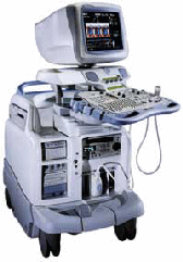
'Vivid 7 Dimension, a premier cardiovascular ultrasound system from GE Healthcare, expands on the strength of a powerful imaging platform to offer new, innovative technology of dimensional proportions.'
Device Information and Specification
CONFIGURATION
Multi-frequency, linear, convex, phased, sector
B-mode, C-mode, M-mode (and 2-D), triplex mode, harmonic imaging, color flow mapping, 3D ultrasound display, power Doppler imaging (PDI), color Doppler, pulsed wave Doppler, continuous wave Doppler, tissue velocity imaging (TVI), tissue type imaging (TTI), strain rate imaging (SRI), tissue synchronization imaging (TSI)
IMAGING OPTIONS
CINE review with 5 speed types, bi- andtri-plane imaging with e.g. stress echo and tissue synchronization imaging
STORAGE, CONNECTIVITY, OS
Patient and image archive, HDD, MOD, DVD, USB flash card, DICOM 3.0 Windows-based
DATA PROCESSING
Digital beamformer with 1024 system processing channel technology
H*W*D m (inch.)
1.58 * 0.64 * 0.89 (62 * 25 * 35)
WEIGHT
191 kg (420 lbs.)
POWER CONSUMPTION
less than 2 KVA
Result Pages : |
Medical-Ultrasound-Imaging.com
former US-TIP.com
Member of SoftWays' Medical Imaging Group - MR-TIP • Radiology TIP • Medical-Ultrasound-Imaging
Copyright © 2008 - 2024 SoftWays. All rights reserved.
Terms of Use | Privacy Policy | Advertise With Us
former US-TIP.com
Member of SoftWays' Medical Imaging Group - MR-TIP • Radiology TIP • Medical-Ultrasound-Imaging
Copyright © 2008 - 2024 SoftWays. All rights reserved.
Terms of Use | Privacy Policy | Advertise With Us
[last update: 2023-11-06 01:42:00]


