Medical Ultrasound Imaging
Sunday, 19 May 2024
Info Sheets Out- side     | 'Gray Scale' p5 Searchterm 'Gray Scale' found in 29 articles 1 term [ • ] - 28 definitions [• ] Result Pages : •
The earliest introduction of vascular ultrasound contrast agents (USCA) was by Gramiak and Shah in 1968, when they injected agitated saline into the ascending aorta and cardiac chambers during echocardiographic to opacify the left heart chamber. Strong echoes were produced within the heart, due to the acoustic mismatch between free air microbubbles in the saline and the surrounding blood.
•
In 1880 the Curie brothers discovered the piezoelectric effect in quartz. Converse piezoelectricity was mathematically deduced from fundamental thermodynamic principles by Lippmann in 1881.
•
In 1917, Paul Langevin (France) and his coworkers developed an underwater sonar system (called hydrophone) that uses the piezoelectric effect to detect submarines through echo location.
•
In 1935, the first RADAR system was produced by the British physicist Robert Watson-Wat. Also about 1935, developments began with the objective to use ultrasonic power therapeutically, utilizing its heating and disruptive effects on living tissues. In 1936, Siemens markets the first ultrasonic therapeutic machine, the Sonostat.
•
Shortly after the World War II, researchers began to explore medical diagnostic capabilities of ultrasound. Karl Theo Dussik (Austria) attempted to locate the cerebral ventricles by measuring the transmission of ultrasound beam through the skull. Other researchers try to use ultrasound to detect gallstones, breast masses, and tumors. These first investigations were performed with A-mode.
•
Shortly after the World War II, researchers in Europe, the United States and Japan began to explore medical diagnostic capabilities of ultrasound. Karl Theo Dussik (Austria) attempted to locate the cerebral ventricles by measuring the transmission of ultrasound beam through the skull. Other researchers, e.g. George Ludwig (United States) tried to use ultrasound to detect gallstones, breast masses, and tumors. This first experimentally investigations were performed with A-mode. Ultrasound pioneers contributed innovations and important discoveries, for example the velocity of sound transmission in animal soft tissues with a mean value of 1540 m/sec (still in use today), and determined values of the optimal scanning frequency of the ultrasound transducer.
•
In the early 50`s the first B-mode images were obtained. Images were static, without gray-scale information in simple black and white and compound technique. Carl Hellmuth Hertz and Inge Edler (Sweden) made in 1953 the first scan of heart activity. Ian Donald and Colleagues (Scotland) were specialized on obstetric and gynecologic ultrasound research. By continuous development it was possible to study pregnancy and diagnose possible complications.
•
After about 1960 two-dimensional compound procedures were developed. The applications in obstetric and gynecologic ultrasound boomed worldwide from the mid 60's with both, A-scan and B-scan equipment. In the late 60's B-mode ultrasonography replaced A-mode in wide parts.
•
In the 70's gray scale imaging became available and with progress of computer technique ultrasonic imaging gets better and faster.
•
•
In the 90's, high resolution scanners with digital beamforming, high transducer frequencies, multi-channel focus and broad-band transducer technology became state of the art. Optimized tissue contrast and improved diagnostic accuracy lead to an important role in breast imaging and cancer detection. Color Doppler and Duplex became available and sensitivity for low flow was continuously improved.
•
Actually, machines with advanced ultrasound system performance are equipped with realtime compound imaging, tissue harmonic imaging, contrast harmonic imaging, vascular assessment, matrix array transducers, pulse inversion imaging, 3D and 4D ultrasound with panoramic view.
•
Imagent® is an injectable suspension for intravenous administration during a sonogram. This diagnostic contrast agent for enhancement of ultrasound images contains perflexane lipid microspheres. Imagent® US is indicated in the assessment of heart function and perfusion, as well as the detection of tumors and blood flow abnormalities by using gray scale, color Doppler, and harmonic ultrasound imaging techniques. During the course of its development, the brand name for this product has changed from Imagent to Imavist (between August 2000 and March 2002) back to Imagent. The manufacturer's 06/03/02 press release announcing FDA approval refers to the product as 'Imagent (formerly Imavist),' and the approval notice and monograph posted at the FDA site refers to the product as Imagent. Jointly developed by Alliance Pharmaceutical Corp and Bayer Schering Pharma AG (Germany). Source: PR Newswire - 10/10/96, 03/31/98, 10/13/99, 03/13/01, 10/08/01; FDA approvals - 05/31/02; Alliance Pharmaceutical press release - 06/03/02. Currently the production of Imagent® is discontinued.
Drug Information and Specification
RESEARCH NAME
AF0150
DEVELOPER
IMCOR Pharmaceuticals, Inc.
INDICATION
APPLICATION
Intravenous
TYPE
Lipid: DMPC
CHARGE
Neutral
Perfluorohexane/Nitrogen
MICROBUBBLE SIZE
99.8% < 10μm
PRESENTATION
-
STORAGE
Room Temp 15−30 °C
PREPARATION
Reconstitute with 10 ml water
DO NOT RELY ON THE INFORMATION PROVIDED HERE, THEY ARE
NOT A SUBSTITUTE FOR THE ACCOMPANYING PACKAGE INSERT! •
The persistence of microbubbles is depended of the shell stability and the density of the gas. This is defined by the equation: (R x d)/(DIFS x constsat) where R is the bubble radius, d the gas density, DIFS the gas diffusivity and constsat the saturation constant. Microbubbles are stabilized with thin coatings of substances such as palmitic acid or by encapsulation in microspheres made with albumin, lipids, or polymers. Low-solubility low-diffusibility gases dramatically improve the persistence. Most recently developed ultrasound contrast agents combine these two approaches to prolong contrast enhancement. Persistence is also a type of temporal smoothing used in both gray scale and color Doppler imaging. Successive frames are averaged as they are displayed to reduce the variations in the image between frames, hence lowering the temporal resolution of the image. •
QB-mode (Quadratic Brightness-mode) images are gray scale images from the quadratic component. QB-mode achieves higher contrast and increased dynamic range than the standard B-mode ultrasound images, without loss in spatial resolution.
Further Reading: Basics:
• 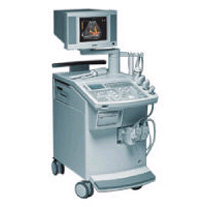 From Siemens Medical Systems;
'This extremely flexible system supports targeted applications for OB/GYN, Radiology, Internal Medicine, Urology, and Prostate Brachytherapy. The large transducer selection, most with high-frequency capabilities, provides the right tool for general imaging, radiology, and internal medicine applications.'
Device Information and Specification
CLINICAL APPLICATION
OB/GYN, cerebrovascular, orthopedics, urology, peripheral vascular, pediatrics, small parts, surgery, abdominal, musculoskeletal
CONFIGURATION
Mobile, compact
DISPLAY MODES
Broadband, high-fidelity, multi-frequency, linear and curved
PROBE PORTS
Two - four
256
MEASUREMENT/CALCULATION FUNCTIONS
OB/GYN measurements and report package, off-line analysis
OPTIONAL PACKAGE
IMAGE PROCESSING
Result Pages : | Share This Page Look Ups |
Medical-Ultrasound-Imaging.com
former US-TIP.com
Member of SoftWays' Medical Imaging Group - MR-TIP • Radiology TIP • Medical-Ultrasound-Imaging
Copyright © 2008 - 2024 SoftWays. All rights reserved.
Terms of Use | Privacy Policy | Advertise With Us
former US-TIP.com
Member of SoftWays' Medical Imaging Group - MR-TIP • Radiology TIP • Medical-Ultrasound-Imaging
Copyright © 2008 - 2024 SoftWays. All rights reserved.
Terms of Use | Privacy Policy | Advertise With Us
[last update: 2023-11-06 01:42:00]