Medical Ultrasound Imaging
Wednesday, 8 May 2024
Info Sheets Out- side     | '4D Ultrasound' p2 Searchterm '4D Ultrasound' found in 15 articles 1 term [ • ] - 10 definitions [• ] - 4 booleans [• ]Result Pages : •
The earliest introduction of vascular ultrasound contrast agents (USCA) was by Gramiak and Shah in 1968, when they injected agitated saline into the ascending aorta and cardiac chambers during echocardiographic to opacify the left heart chamber. Strong echoes were produced within the heart, due to the acoustic mismatch between free air microbubbles in the saline and the surrounding blood.
•
In 1880 the Curie brothers discovered the piezoelectric effect in quartz. Converse piezoelectricity was mathematically deduced from fundamental thermodynamic principles by Lippmann in 1881.
•
In 1917, Paul Langevin (France) and his coworkers developed an underwater sonar system (called hydrophone) that uses the piezoelectric effect to detect submarines through echo location.
•
In 1935, the first RADAR system was produced by the British physicist Robert Watson-Wat. Also about 1935, developments began with the objective to use ultrasonic power therapeutically, utilizing its heating and disruptive effects on living tissues. In 1936, Siemens markets the first ultrasonic therapeutic machine, the Sonostat.
•
Shortly after the World War II, researchers began to explore medical diagnostic capabilities of ultrasound. Karl Theo Dussik (Austria) attempted to locate the cerebral ventricles by measuring the transmission of ultrasound beam through the skull. Other researchers try to use ultrasound to detect gallstones, breast masses, and tumors. These first investigations were performed with A-mode.
•
Shortly after the World War II, researchers in Europe, the United States and Japan began to explore medical diagnostic capabilities of ultrasound. Karl Theo Dussik (Austria) attempted to locate the cerebral ventricles by measuring the transmission of ultrasound beam through the skull. Other researchers, e.g. George Ludwig (United States) tried to use ultrasound to detect gallstones, breast masses, and tumors. This first experimentally investigations were performed with A-mode. Ultrasound pioneers contributed innovations and important discoveries, for example the velocity of sound transmission in animal soft tissues with a mean value of 1540 m/sec (still in use today), and determined values of the optimal scanning frequency of the ultrasound transducer.
•
In the early 50`s the first B-mode images were obtained. Images were static, without gray-scale information in simple black and white and compound technique. Carl Hellmuth Hertz and Inge Edler (Sweden) made in 1953 the first scan of heart activity. Ian Donald and Colleagues (Scotland) were specialized on obstetric and gynecologic ultrasound research. By continuous development it was possible to study pregnancy and diagnose possible complications.
•
After about 1960 two-dimensional compound procedures were developed. The applications in obstetric and gynecologic ultrasound boomed worldwide from the mid 60's with both, A-scan and B-scan equipment. In the late 60's B-mode ultrasonography replaced A-mode in wide parts.
•
In the 70's gray scale imaging became available and with progress of computer technique ultrasonic imaging gets better and faster.
•
•
In the 90's, high resolution scanners with digital beamforming, high transducer frequencies, multi-channel focus and broad-band transducer technology became state of the art. Optimized tissue contrast and improved diagnostic accuracy lead to an important role in breast imaging and cancer detection. Color Doppler and Duplex became available and sensitivity for low flow was continuously improved.
•
Actually, machines with advanced ultrasound system performance are equipped with realtime compound imaging, tissue harmonic imaging, contrast harmonic imaging, vascular assessment, matrix array transducers, pulse inversion imaging, 3D and 4D ultrasound with panoramic view.
• 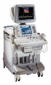 From GE Healthcare.;
'The System of Choice for Shared Service. The LOGIQ® 7 system provides a full range of clinical applications including abdominal, small parts, surgery, vascular and cardiac imaging and the power of GE's patented TruScan architecture. Just imagine an ultrasound system so versatile and reliable that it can meet the demands of virtually any clinical setting. And an ergonomic design that improves scanning comfort and clinical work flow.'
Device Information and Specification
APPLICATIONS
Abdominal, cardiac, breast, intraoperative, musculoskeletal, neonatal, OB/GYN, orthopedic, pediatric, small parts, transcranial, urologic, vascular
CONFIGURATION
17' high resolution non-interlaced flat CRT, 4 active probe ports
B-mode, M-mode, coded harmonic imaging, color flow mode (CFM), power Doppler imaging (PDI), color Doppler, pulsed wave Doppler, tissue harmonic imaging
IMAGING OPTIONS
CrossXBeam spatial compounding, coded ultrasound acquisition),speckle reduction imaging (SRI), TruScan technology store raw data, CINE review with 4 speed types
OPTIONAL PACKAGE
Transesophageal scanning, stress echo, tissue velocity imaging (TVI), tissue velocity Doppler (TVD), contrast harmonic imaging
STORAGE, CONNECTIVITY, OS
Patient and image archive, HDD, DICOM 3.0, CD/DVD, MOD, Windows-based
DATA PROCESSING
Digital beamformer with 1024 system processing channel technology
H*W*D m (inch.)
1.62 * 0.61 * 0.99 (64 * 24 * 39)
WEIGHT
246 kg (498 lbs.)
POWER CONSUMPTION
less than 1.5 KVA
• 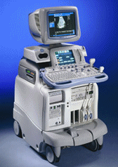 From GE Healthcare.;
'The System of Choice for General Imaging Imagine a leading-edge ultrasound system so versatile that it can meet the demands of virtually any clinical setting. With the LOGIQ® 9, you'll have a high-performance system capable of multi-dimensional imaging for a full range of clinical applications - from abdominal to breast to vascular imaging. And an ergonomic design that improves scanning comfort and clinical work flow. Now, imagine what LOGIQ® 9 could do for you and your patients.'
Device Information and Specification
APPLICATIONS
Abdominal, cardiac, breast, intraoperative, musculoskeletal, neonatal, OB/GYN, orthopedic, pediatric, small parts, transcranial, urologic, vascular
CONFIGURATION
17' high resolution non-interlaced flat CRT, 4 active probe ports
B-mode, M-mode, coded harmonic imaging, color flow mode (CFM), power Doppler imaging (PDI), PW-HPRF, CW Doppler, color Doppler, pulsed wave Doppler, tissue harmonic imaging
IMAGING OPTIONS
CrossXBeam spatial compounding, coded ultrasound acquisition), speckle reduction imaging (SRI), TruScan technology store raw data, real-time 4D ultrasound, Tru 3D ultrasound
STORAGE, CONNECTIVITY, OS
Patient and image archive, HDD, DICOM 3.0, CD/DVD, MOD, PCMCIA, USB, Windows-based
DATA PROCESSING
Digital beamformer with 1024 system processing channel technology
H*W*D m (inch.)
1.62 * 0.61 * 0.99 (64 * 24 * 39)
WEIGHT
202 kg (408 lb.)
POWER CONSUMPTION
less than 2 KVA
•
The definition of imaging is the visual representation of an object. Medical imaging is a broad term that encompasses various imaging modalities and techniques used in the field of medicine to visualize and study the body's anatomy and physiology. It includes both diagnostic and non-diagnostic imaging procedures, where diagnostic imaging specifically refers to the subset of medical imaging techniques that are primarily focused on diagnosing diseases or conditions. Medical imaging techniques are employed to obtain images or visual representations of the internal organs, tissues, and structures, aiding in the diagnosis, treatment, and monitoring of medical conditions.
The field of medical imaging has significantly evolved since the discovery of X-rays by Konrad Roentgen in 1896. Initially, radiological imaging involved focusing X-rays on the body and capturing the images on a single piece of film within a specialized cassette. Subsequent advancements introduced the use of fluorescent screens and special glasses for real-time visualization of X-ray images. A significant breakthrough came with the application of contrast agents, enhancing image contrast and improving organ visualization. In the 1950s, nuclear medicine studies utilizing gamma cameras demonstrated the uptake of low-level radioactive chemicals in organs, enabling the observation of biological processes in vivo. Currently, positron emission tomography (PET) and single photon emission computed tomography (SPECT) technologies play pivotal roles in clinical research and the diagnosis of biochemical and physiological processes. Additionally, the advent of the x-ray image intensifier in 1955 facilitated the capture and display of x-ray movies. In the 1960s, diagnostic imaging incorporated the principles of sonar, using ultrasonic waves generated by a quartz crystal. These waves, reflecting at the interfaces between different tissues, were received by ultrasound machines and translated into images through computer algorithms and reconstruction software. Ultrasound (ultrasonography) has become an indispensable diagnostic tool across various medical specialties, with immense potential for further advancements such as targeted contrast imaging, real-time 3D or 4D ultrasound, and molecular imaging. The first use of ultrasound contrast agents (USCA) dates back to 1968. Digital imaging techniques were introduced in the 1970s, revolutionizing conventional fluoroscopic image intensifiers. Godfrey Hounsfield's pioneering work led to the development of the first computed tomography (CT) scanner. Digital images are now electronic snapshots represented as grids of dots or pixels. X-ray CT brought about a breakthrough in medical imaging by providing cross-sectional images of the human body with high contrast between different types of soft tissue. These advancements were made possible by analog-to-digital converters and computers. The introduction of multislice spiral CT technology dramatically expanded the clinical applications of CT scans. The first magnetic resonance imaging (MRI) devices were tested on clinical patients in 1980. With technological improvements, such as higher field strength, more open MRI magnets, faster gradient systems, and novel data-acquisition techniques, MRI has emerged as a real-time interactive imaging modality capable of providing detailed structural and functional information of the body. Today, imaging in medicine offers a wide range of modalities, including:
•
X-ray projection imaging;
•
Fluoroscopy;
•
Computed tomography (CT / CAT);
•
Ultrasound imaging (US)
•
Magnetic resonance imaging (MRI), Magnetic source imaging (MSI);
•
Single photon emission computed tomography (SPECT);
•
Positron emission tomography (PET);
•
Mammography.
These imaging modalities have become integral components of modern healthcare. With the rapid advancement of digital imaging, efficient management has become important, leading to the expansion of radiology information systems (RIS) and the adoption of Picture Archiving and Communication Systems (PACS) for digital image archiving. In telemedicine, real-time transmission of all medical image modalities from MRI to X-ray, CT and ultrasound has become the standard. The field of medical imaging continues to evolve, promising further innovations and advancements in the future, ultimately contributing to improved patient care and diagnostics. See also History of Ultrasound Contrast Agents, and History of Ultrasound. Further Reading: News & More:
•
Ultrasound is the ideal tool to examine children of all ages. It is fast, painless, uses no ionizing radiation, and does not require a baby to remain still for long periods. Real-time modes show movement of internal tissues and organs. Advanced ultrasound imaging techniques such as color Doppler, 4D ultrasound, harmonic imaging, and higher resolution, as well as the application of ultrasound contrast agents broaden the potential of ultrasound. Pediatric [paediatric, Brit.] ultrasound can be used in all body regions and reduce the number of more invasive or radiating examinations that often additionally need sedation or intravenous iodinated contrast agents. See also Fetal Ultrasound, Reflux Sonography, Ultrasound Safety, Abdominal Ultrasound and Pregnancy Ultrasound. Result Pages : | Share This Page Look Ups |
Medical-Ultrasound-Imaging.com
former US-TIP.com
Member of SoftWays' Medical Imaging Group - MR-TIP • Radiology TIP • Medical-Ultrasound-Imaging
Copyright © 2008 - 2024 SoftWays. All rights reserved.
Terms of Use | Privacy Policy | Advertise With Us
former US-TIP.com
Member of SoftWays' Medical Imaging Group - MR-TIP • Radiology TIP • Medical-Ultrasound-Imaging
Copyright © 2008 - 2024 SoftWays. All rights reserved.
Terms of Use | Privacy Policy | Advertise With Us
[last update: 2023-11-06 01:42:00]