Medical Ultrasound Imaging
Sunday, 19 May 2024
Info Sheets Out- side     | 'B-Mode' p2 Searchterm 'B-Mode' found in 58 articles 6 terms [ • ] - 52 definitions [• ] Result Pages : •
(BAT) B-mode acquisition and targeting is a stereotactic tumor locating device system, based on ultrasound and designed to maximize the precision of external beam radiation. By tracking the position and orientation of the ultrasound transceiver within the treatment vault, BAT ultrasound produces images that can be fused with the CT scans used in the treatment plan. When this is done directly before treatment, the current location of the target volume can be determined in reference to the treatment plan so that the volume is placed precisely and accurately. The patient's position can then be adjusted accordingly. •
Duplex ultrasonography (duplex scan) consists of two ultrasound modalities to study blood flow and the perivascular tissue. This includes B-mode / gray scale imaging used in combination with spectral Doppler / pulsed-wave Doppler. The real-time visualization of the vessels and tissue by the B-mode component improves the PW Doppler positioning and the direction of blood flow can be inferred. The angle between the direction of the PW Doppler signal and the estimated direction of blood flow can be measured. Duplex techniques are available on phased array, linear array, and mechanical scanners. A phased array probe is able to create nearly simultaneous images and flow information. A linear array transducer can also do this if the Doppler probe is attached separately to one end of the scanhead. A mechanical transducer freeze the image; the crystals must be static to produce a Doppler image. The first two transducers are therefore the best choice for Duplex. See also Compound B-Mode, and Duplex Scanner. Further Reading: News & More:
•
The earliest introduction of vascular ultrasound contrast agents (USCA) was by Gramiak and Shah in 1968, when they injected agitated saline into the ascending aorta and cardiac chambers during echocardiographic to opacify the left heart chamber. Strong echoes were produced within the heart, due to the acoustic mismatch between free air microbubbles in the saline and the surrounding blood.
•
In 1880 the Curie brothers discovered the piezoelectric effect in quartz. Converse piezoelectricity was mathematically deduced from fundamental thermodynamic principles by Lippmann in 1881.
•
In 1917, Paul Langevin (France) and his coworkers developed an underwater sonar system (called hydrophone) that uses the piezoelectric effect to detect submarines through echo location.
•
In 1935, the first RADAR system was produced by the British physicist Robert Watson-Wat. Also about 1935, developments began with the objective to use ultrasonic power therapeutically, utilizing its heating and disruptive effects on living tissues. In 1936, Siemens markets the first ultrasonic therapeutic machine, the Sonostat.
•
Shortly after the World War II, researchers began to explore medical diagnostic capabilities of ultrasound. Karl Theo Dussik (Austria) attempted to locate the cerebral ventricles by measuring the transmission of ultrasound beam through the skull. Other researchers try to use ultrasound to detect gallstones, breast masses, and tumors. These first investigations were performed with A-mode.
•
Shortly after the World War II, researchers in Europe, the United States and Japan began to explore medical diagnostic capabilities of ultrasound. Karl Theo Dussik (Austria) attempted to locate the cerebral ventricles by measuring the transmission of ultrasound beam through the skull. Other researchers, e.g. George Ludwig (United States) tried to use ultrasound to detect gallstones, breast masses, and tumors. This first experimentally investigations were performed with A-mode. Ultrasound pioneers contributed innovations and important discoveries, for example the velocity of sound transmission in animal soft tissues with a mean value of 1540 m/sec (still in use today), and determined values of the optimal scanning frequency of the ultrasound transducer.
•
In the early 50`s the first B-mode images were obtained. Images were static, without gray-scale information in simple black and white and compound technique. Carl Hellmuth Hertz and Inge Edler (Sweden) made in 1953 the first scan of heart activity. Ian Donald and Colleagues (Scotland) were specialized on obstetric and gynecologic ultrasound research. By continuous development it was possible to study pregnancy and diagnose possible complications.
•
After about 1960 two-dimensional compound procedures were developed. The applications in obstetric and gynecologic ultrasound boomed worldwide from the mid 60's with both, A-scan and B-scan equipment. In the late 60's B-mode ultrasonography replaced A-mode in wide parts.
•
In the 70's gray scale imaging became available and with progress of computer technique ultrasonic imaging gets better and faster.
•
•
In the 90's, high resolution scanners with digital beamforming, high transducer frequencies, multi-channel focus and broad-band transducer technology became state of the art. Optimized tissue contrast and improved diagnostic accuracy lead to an important role in breast imaging and cancer detection. Color Doppler and Duplex became available and sensitivity for low flow was continuously improved.
•
Actually, machines with advanced ultrasound system performance are equipped with realtime compound imaging, tissue harmonic imaging, contrast harmonic imaging, vascular assessment, matrix array transducers, pulse inversion imaging, 3D and 4D ultrasound with panoramic view.
• 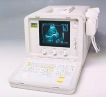 From SIUI Inc.;
'The CTS-385 Plus is designed for the diagnosis of liver, gallbladder, kidney, pancreas, thyroid, breast, uterus, bladder, ovary, etc. The system is a portable linear and convex unit for general application.' Features: 'High quality image Cineloop − 32-frame non-volatile storage capacity Probe frequency conversation option Computer image communication Various measuring function Foldaway keyboard for easy operation Dual probe connector'
Device Information and Specification
APPLICATIONS
See description above
CONFIGURATION
Portable, gray scale(256)
Linear and convex
PROBES STANDARD
1 * 2.5MHz ~ 5.0MHz trifrequency convex probe
2.5MHz to 10.0MHz, linear and convex, broad band, trifrequency
IMAGING OPTIONS
OPTIONAL PACKAGE
DATA PROCESSING
Pre-processing, correlation-processing, interpolation
H*W*D m
0.26 * 0.3 * 0.41
WEIGHT
10 - 13 kg
POWER REQUIREMENT
AC 220V/110V, 50Hz/60Hz
POWER CONSUMPTION
0.1 KVA
• 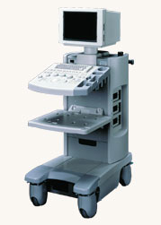 From Hitachi Medical Systems America Inc.;
The EUB-2000 B/W is a compact system capable of B-mode, M-mode, and optional with grayscale Doppler (EUB-2000 B/W Dop). This unit features intuitive operation, high mobility, and exceptional image quality. When color Doppler is not required, this system is an excellent choice for general purpose scanning. Device Information and Specification
APPLICATIONS
General purpose, OB/GYN, EUS, brachytherapy
CONFIGURATION
Compact system
Triple frequency
Linear, convex, radial, bi-plane, echoendoscope longitudinal
PROBE PORTS
Two
IMAGING OPTIONS
Simultaneous imaging with biplane probes
ADDITIONAL FEATURES
Cine / multi memory, application specific measuring, etc.
Result Pages : | Share This Page Look Ups |
Medical-Ultrasound-Imaging.com
former US-TIP.com
Member of SoftWays' Medical Imaging Group - MR-TIP • Radiology TIP • Medical-Ultrasound-Imaging
Copyright © 2008 - 2024 SoftWays. All rights reserved.
Terms of Use | Privacy Policy | Advertise With Us
former US-TIP.com
Member of SoftWays' Medical Imaging Group - MR-TIP • Radiology TIP • Medical-Ultrasound-Imaging
Copyright © 2008 - 2024 SoftWays. All rights reserved.
Terms of Use | Privacy Policy | Advertise With Us
[last update: 2023-11-06 01:42:00]