Medical Ultrasound Imaging
Tuesday, 21 May 2024
Info Sheets Out- side     | 'Real Time' p2 Searchterm 'Real Time' found in 22 articles 5 definitions [ • ] - 17 booleans [• ]Result Pages : •
Real-time mode has been developed to present motion like a movie of the body's inner workings, showing this information at a high rate. The special real-time transducer uses a larger sound beam than for A, B or M-modes. A linear array transducer with multiple crystal elements displays real-time compound B-mode images with up to 100 images per second. At each scan line, one sound pulse is transmitted and all echoes from the surface to the deepest range are received. Then the ultrasound beam moves on to the next scan line position where pulse transmission and echo recording are repeated. See also Compound B-Mode, Pulse Inversion Doppler, and Frame Averaging. Further Reading: News & More:
•
Most usual ultrasound machines are 2D real-time systems. This types of ultrasound scanners allow to assess both motion and anatomy, including the motion of heart valves, the movement of intestines and lungs and also to guide interventions, like for example a biopsy or a laparoscopic ultrasound. A standard real-time scanner consists of a mobile console with the monitor on the top and rows of small containers at the bottom to accommodate a variety of scanner probes. The linear, curved or phased array transducers are usually equipped with multiple crystals or in some cases with a moving crystal. A real-time scanner may be e.g., a mechanical scanner or electronic array scanner. See also Musculoskeletal and Joint Ultrasound. Further Reading: News & More: •
Transducers used for the real-time mode are different than for the A-mode, B-, or M-modes. A linear array transducer with multiple piezoelectric crystal elements that are different arranged and fired, transmits the needed larger sound beam. A subgroup of x adjacent elements (8-16; or more in wide-aperture designs) is pulsed simultaneously; the inner elements pulse delayed with respect to the outer elements. The interference of the x small divergent wavelets generates a focused beam. The delay time determining the focus depth of a real-time transducer can be changed during imaging. Similar delay factors applied during the receiving phase, result in a dynamic focusing effect on the return. This forms a single scan line in the real-time image. To produce the following scan line, another group of x elements is selected by shifting one element position along the transducer array from the previous group. This pattern is then repeated for the groups along the array, in a sequential and repetitive way. Further Reading: Basics: • 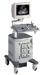 From ALOKA Co., Ltd.;
'A Platform for Digital, Pure-Beam Imaging The high-performance, ALOKA ProSound SSD-3500 utilizes advanced ProSound technologies including: Fully digital beam former A wide dynamic range, 12-bit A/D converter Multi beam processing. The SSD-3500 also helps you achieve more efficient examinations. Its ergonomic, user-friendly design enables you to customize the system according to your specific application needs.'
Device Information and Specification
APPLICATIONS
CONFIGURATION
Compact, portable, dual dynamic display
Color Flow, Power Flow, Spectral Doppler, Real-time Free Angular M-Mode, Tissue Harmonic Imaging, Quint Frequency Imaging, Pure Harmonic Detection
STORAGE, CONNECTIVITY, OS
Data Management Subsystem (iDMS), DICOM-Worklist
DATA PROCESSING
12-bit analog to digital converter
•  From GE Healthcare.;
'GE is defining a new age of ultrasound. We call it Volume Ultrasound. GE's Voluson 730 Expert is a powerful system that enables real-time techniques for acquiring, navigating and analyzing volumetric images so that you can make clinical decisions with unprecedented confidence.'
Device Information and Specification
APPLICATIONS
Abdominal, breast, cardiac, musculoskeletal, neonatal, OB/GYN, pediatric, small parts, transcranial, urological, vascular
CONFIGURATION
15' high resolution non-interlaced flat CRT, 4 active probe ports
B-mode, M-mode, coded harmonic imaging (2-D), color flow mode (CFM), power Doppler imaging (PDI), color Doppler, pulsed wave Doppler, high pulse repetition frequency (HPRF) Doppler, tissue harmonic imaging, 3-D power Doppler
IMAGING OPTIONS
CrossXBeam spatial compounding, coded excitation , spatio-temporal image correlation (STIC), B-Flow (simultaneous imaging of tissue and blood flow), strain rate imaging (SRI)
OPTIONAL PACKAGE
STORAGE, CONNECTIVITY, OS
SonoView archiving and data management, network, HDD, DICOM 3.0, CD/DVD, MOD, USB, Windows-based
DATA PROCESSING
Digital beamformer with 512 system processing channel technology
H*W*D m (inch.)
1.43 * 0.69 * 1.02 (56 * 27 * 40)
WEIGHT
136 kg (300 lbs.)
Result Pages : | Share This Page Look Ups |
Medical-Ultrasound-Imaging.com
former US-TIP.com
Member of SoftWays' Medical Imaging Group - MR-TIP • Radiology TIP • Medical-Ultrasound-Imaging
Copyright © 2008 - 2024 SoftWays. All rights reserved.
Terms of Use | Privacy Policy | Advertise With Us
former US-TIP.com
Member of SoftWays' Medical Imaging Group - MR-TIP • Radiology TIP • Medical-Ultrasound-Imaging
Copyright © 2008 - 2024 SoftWays. All rights reserved.
Terms of Use | Privacy Policy | Advertise With Us
[last update: 2023-11-06 01:42:00]