Medical Ultrasound Imaging
Sunday, 12 May 2024
Info Sheets Out- side     | 'Real' p2 Searchterm 'Real' found in 60 articles 3 terms [ • ] - 57 definitions [• ] Result Pages : •  From Hitachi Medical Corporation (HMC);
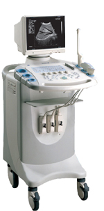
The HI VISION™ 6500 - EUB-6500 high resolution digital ultrasound system offers advanced clinical imaging, enhanced operating efficiency, and remarkable clinical flexibility, all in robust and versatile configuration that simply represents a better clinical solution in a variety of real-world, real-work arenas.
Device Information and Specification
APPLICATIONS
Abdominal, brachytherapy/cryotherapy, breast, cardiac, dedicated biopsy, endoscopic, intraoperative, laparoscopic, musculoskeletal, OB/GYN, pediatric, small parts, urologic, vascular
CONFIGURATION
Compact system
Linear, convex, radial, miniradial/miniprobe, biplane, phased array, echoendoscope longitudinal, echoendoscope radial
Tissue Doppler imaging (TDI), pulsed wave Doppler, continuous wave Doppler, color flow imaging, power Doppler, directional power Doppler, color flow angiography, real-time Doppler measurements, 4 modes of dynamic tissue harmonic imaging (dTHI), wideband pulse inversion imaging (WPI)
IMAGING OPTIONS
3RD generation color artifact suppression
OPTIONAL PACKAGE
3D ultrasound, dual omni-directional M-mode display, steerable CW Doppler, dynamic contrast harmonics imaging, stress echo, Pentax EUS and Fujinon Mini-probe
STORAGE, CONNECTIVITY, OS
Patient and image database management system, HDD, FDD, MOD, CD-ROM, Network, DICOM 3.0, Windows XP
DATA PROCESSING
H*W*D m (inch.)
1.40 x 0.51 x 0.79 (55 x 20 x 31)
WEIGHT
130 kg (286 lbs.)
POWER CONSUMPTION
1.2kVA
ENVIRONMENTAL POLLUTION
4096 btu/hr heat output
•
Interventional ultrasound, also known as ultrasonography, encompasses a range of invasive or surgical procedures guided by ultrasound imaging. While its widest application lies in intravascular ultrasound imaging for measuring atherosclerotic plaque, it has proven valuable in various medical fields. In urology, ultrasound-guided interventions are employed for treatments like high intensity focused ultrasound (HIFU) in prostate conditions. The precise imaging provided by ultrasound aids in targeting the affected area and delivering therapeutic energy effectively. In intraabdominal conditions, endoscopic ultrasound is frequently utilized. This technique combines ultrasound imaging with an endoscope to visualize and evaluate structures within the gastrointestinal tract, allowing for precise diagnoses and targeted interventions. Ultrasound-guided procedures play a significant role in several medical specialties, including liver sonography, obstetric and gynecologic ultrasound, and thyroid ultrasound. These procedures involve interventions such as RF thermal ablation or biopsies, which are guided by real-time ultrasound imaging. For instance, in liver sonography, ultrasound guidance is crucial for performing biopsies or RF thermal ablation, a technique used to treat liver tumors by delivering localized heat to destroy the abnormal tissue. The real-time imaging allows for precise needle placement and monitoring during the procedure. In obstetric and gynecologic ultrasound, ultrasound-guided procedures, such as biopsies, can be performed to obtain tissue samples for diagnostic purposes. Additionally, ultrasound guidance is valuable during interventions like amniocentesis or fetal blood sampling, enabling accurate and safe procedures. Thyroid ultrasound procedures often involve ultrasound-guided fine-needle aspiration biopsy (FNAB), which allows for the sampling of thyroid nodules for cytological examination. The ultrasound image helps guide the needle into the targeted area, ensuring accurate sampling and minimizing potential complications. Overall, ultrasound-guided interventions provide minimally invasive and precise approaches to diagnosis and treatment. The real-time imaging capabilities of ultrasound contribute to enhanced accuracy, safety, and patient outcomes in procedures like biopsies, injections, and drainage. See also Transurethral Sonography, Endocavitary Echography, and B-Mode Acquisition and Targeting. •  From SIUI Inc.;
'Dedicated to ultrasound industry, Shantou Institute of Ultrasonic Instruments, Inc. (SIUI) has launched Apogee 3500, the Digital Color Doppler Ultrasound Imaging System. With latest imaging technologies, high-definition image quality and excellent practical functions, the Apogee 3500 offers optimal solutions for clinical ultrasonic examination.' 'The Apogee 3500 is available with many high-density, super broadband and multi-frequency probes, such as convex, micro-convex, linear, vaginal, rectal and phased array probes, which are widely applied for different clinical diagnoses, including abdomen (liver, kidney, gall-bladder, pancreas), gynecology (uterus, ovary), obstetrics (early pregnancy, basic OB, complete OB, multi gestation, fetal echo), cardiology (adult and pediatric cardiology), small parts (thyroid, galactophore, testicles, neonate), peripheral vascular and prostate.'
Device Information and Specification
CONFIGURATION
Normal system, color - gray scale(256)
Linear, convex and phased array
PROBES STANDARD
2.0 MHz ~ 12.0 MHz, broad band, tri-frequency
B-mode (B, 2B, 4B), M-mode, B/M-mode, real-time compound imaging, panoramic imaging, trapezoidal imaging (linear probes), spectrum Doppler (PWD and CWD), color Doppler flow imaging (CDFI), color power angio (CPA), tissue harmonic imaging (THI)
IMAGING OPTIONS
Real-time ZOOM, zoom rate and position selectable
OPTIONAL PACKAGE
H*W*D m
1.29 * 0.52 * 0.75
WEIGHT
110 kg
POWER REQUIREMENT
AC 220V/110V, 50Hz/60Hz
POWER CONSUMPTION
0.6 KVA
•
Also called B-mode echography, B-mode sonography, 2D-mode, and sonogram. B-mode ultrasound (Brightness-mode) is the display of a 2D-map of B-mode data, currently the most common form of ultrasound imaging. The development from A-mode to B-mode is that the ultrasound signal is used to produce various points whose brightness depends on the amplitude instead of the spiking vertical movements in the A-mode. Sweeping a narrow ultrasound beam through the area being examined while transmitting pulses and detecting echoes along closely spaced scan lines produces B-scan images. The vertical position of each bright dot is determined by the time delay from pulse transmission to return of the echo, and the horizontal position by the location of the receiving transducer element. To generate a rapid series of individual 2D images that show motion, the ultrasound beam is swept repeatedly. The returning sound pulses in B-mode have different shades of darkness depending on their intensities. The varying shades of gray reflect variations in the texture of internal organs. This form of display (solid areas appear white and fluid areas appear black) is also called gray scale. Different types of displayed B-mode images are: The probe movement can be performed manual (compound and static B-scanner) or automatic (real-time scanner). The image reconstruction can be parallel or sector type. See also B-Scan, 4B-Mode, and Harmonic B-Mode Imaging. Further Reading: News & More:
• 
From SIUI Inc.;
Device Information and Specification
CONFIGURATION
Normal system, gray scale(256)
Linear and convex
PROBES STANDARD
2.5MHz ~ 11.0MHz, broad band, tri-frequency
IMAGING OPTIONS
Real zoom, max. Zoom * 4.0, multiplying power and position selectable
OPTIONAL PACKAGE
H*W*D m
1.30 * 0.52 * 0.77
WEIGHT
90kg (main unit)
POWER REQUIREMENT
AC 220V/110V, 50Hz/60Hz
POWER CONSUMPTION
0.45 KVA
Result Pages : | Share This Page Look Ups |
Medical-Ultrasound-Imaging.com
former US-TIP.com
Member of SoftWays' Medical Imaging Group - MR-TIP • Radiology TIP • Medical-Ultrasound-Imaging
Copyright © 2008 - 2024 SoftWays. All rights reserved.
Terms of Use | Privacy Policy | Advertise With Us
former US-TIP.com
Member of SoftWays' Medical Imaging Group - MR-TIP • Radiology TIP • Medical-Ultrasound-Imaging
Copyright © 2008 - 2024 SoftWays. All rights reserved.
Terms of Use | Privacy Policy | Advertise With Us
[last update: 2023-11-06 01:42:00]